3KCG

Crystal structure of the antithrombin-factor IXa-pentasaccharide complex


Experimental Data Snapshot

  • Method: X-RAY DIFFRACTION
  • Resolution: 1.70 Å
  • R-Value Free: 0.230 
  • R-Value Work: 0.208 
  • R-Value Observed: 0.208 

wwPDB Validation   3D Report Full Report


This is version 2.2 of the entry. See complete history


Literature

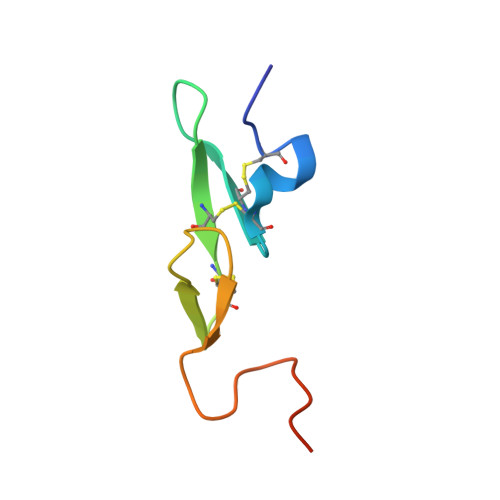
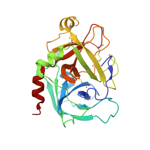
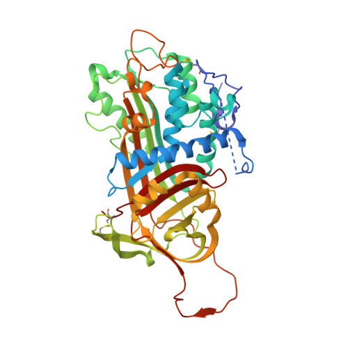
Molecular basis of factor IXa recognition by heparin-activated antithrombin revealed by a 1.7-A structure of the ternary complex.

Johnson, D.J.Langdown, J.Huntington, J.A.

(2010) Proc Natl Acad Sci U S A 107: 645-650

  • DOI: https://doi.org/10.1073/pnas.0910144107
  • Primary Citation of Related Structures:  
    3KCG

  • PubMed Abstract: 

    Factor (f) IXa is a critical enzyme for the formation of stable blood clots, and its deficiency results in hemophilia. The enzyme functions at the confluence of the intrinsic and extrinsic pathways by binding to fVIIIa and rapidly generating fXa. In spite of its importance, little is known about how fIXa recognizes its cofactor, its substrate, or its only known inhibitor, antithrombin (AT). However, it is clear that fIXa requires extensive exosite interactions to present substrates for efficient cleavage. Here we describe the 1.7-A crystal structure of fIXa in its recognition (Michaelis) complex with heparin-activated AT. It represents the highest resolution structure of both proteins and allows us to address several outstanding issues. The structure reveals why the heparin-induced conformational change in AT is required to permit simultaneous active-site and exosite interactions with fIXa and the nature of these interactions. The reactive center loop of AT has evolved to specifically inhibit fIXa, with a P2 Gly so as not to clash with Tyr99 on fIXa, a P4 Ile to fit snugly into the S4 pocket, and a C-terminal extension to exploit a unique wall-like feature of the active-site cleft. Arg150 is at the center of the exosite interface, interacting with AT residues on beta-sheet C. A surprising crystal contact is observed between the heparin pentasaccharide and fIXa, revealing a plausible mode of binding that would allow longer heparin chains to bridge the complex.


  • Organizational Affiliation

    Department of Haematology, Cambridge Institute for Medical Research, University of Cambridge, Cambridge CB2 0XY, United Kingdom.


Macromolecules
Find similar proteins by:  (by identity cutoff)  |  3D Structure
Entity ID: 1
MoleculeChains Sequence LengthOrganismDetailsImage
Coagulation factor IXa light chainA [auth L]59Homo sapiensMutation(s): 0 
Gene Names: F9
EC: 3.4.21.22
UniProt & NIH Common Fund Data Resources
Find proteins for P00740 (Homo sapiens)
Explore P00740 
Go to UniProtKB:  P00740
PHAROS:  P00740
GTEx:  ENSG00000101981 
Entity Groups  
Sequence Clusters30% Identity50% Identity70% Identity90% Identity95% Identity100% Identity
UniProt GroupP00740
Sequence Annotations
Expand
  • Reference Sequence
Find similar proteins by:  (by identity cutoff)  |  3D Structure
Entity ID: 2
MoleculeChains Sequence LengthOrganismDetailsImage
Coagulation factor IXa heavy chainB [auth H]235Homo sapiensMutation(s): 1 
Gene Names: F9
EC: 3.4.21.22
UniProt & NIH Common Fund Data Resources
Find proteins for P00740 (Homo sapiens)
Explore P00740 
Go to UniProtKB:  P00740
PHAROS:  P00740
GTEx:  ENSG00000101981 
Entity Groups  
Sequence Clusters30% Identity50% Identity70% Identity90% Identity95% Identity100% Identity
UniProt GroupP00740
Sequence Annotations
Expand
  • Reference Sequence
Find similar proteins by:  (by identity cutoff)  |  3D Structure
Entity ID: 3
MoleculeChains Sequence LengthOrganismDetailsImage
Antithrombin-IIIC [auth I]432Homo sapiensMutation(s): 1 
Gene Names: SERPINC1AT3PRO0309
UniProt & NIH Common Fund Data Resources
Find proteins for P01008 (Homo sapiens)
Explore P01008 
Go to UniProtKB:  P01008
PHAROS:  P01008
GTEx:  ENSG00000117601 
Entity Groups  
Sequence Clusters30% Identity50% Identity70% Identity90% Identity95% Identity100% Identity
UniProt GroupP01008
Sequence Annotations
Expand
  • Reference Sequence
Oligosaccharides

Help

Entity ID: 4
MoleculeChains Length2D Diagram Glycosylation3D Interactions
alpha-L-fucopyranose-(1-6)-2-acetamido-2-deoxy-beta-D-glucopyranoseD [auth A],
F [auth C]
2N-Glycosylation
Glycosylation Resources
GlyTouCan:  G86851RC
GlyCosmos:  G86851RC
GlyGen:  G86851RC
Entity ID: 5
MoleculeChains Length2D Diagram Glycosylation3D Interactions
alpha-D-mannopyranose-(1-4)-2-acetamido-2-deoxy-beta-D-glucopyranose-(1-4)-2-acetamido-2-deoxy-beta-D-glucopyranoseE [auth B]3N-Glycosylation
Glycosylation Resources
GlyTouCan:  G62182OO
GlyCosmos:  G62182OO
GlyGen:  G62182OO
Entity ID: 6
MoleculeChains Length2D Diagram Glycosylation3D Interactions
3,4-di-O-methyl-2,6-di-O-sulfo-alpha-D-glucopyranose-(1-4)-2,3-di-O-methyl-beta-D-glucopyranuronic acid-(1-4)-2,3,6-tri-O-sulfo-alpha-D-glucopyranose-(1-4)-3-O-methyl-2-O-sulfo-alpha-L-idopyranuronic acid-(1-4)-methyl 2,3,6-tri-O-sulfo-alpha-D-glucopyranosideG [auth D]5N/A
Glycosylation Resources
GlyTouCan:  G76630SM
GlyCosmos:  G76630SM
Biologically Interesting Molecules (External Reference) 1 Unique
Experimental Data & Validation

Experimental Data

  • Method: X-RAY DIFFRACTION
  • Resolution: 1.70 Å
  • R-Value Free: 0.230 
  • R-Value Work: 0.208 
  • R-Value Observed: 0.208 
  • Space Group: P 21 21 21
Unit Cell:
Length ( Å )Angle ( ˚ )
a = 78.78α = 90
b = 88.44β = 90
c = 147.23γ = 90
Software Package:
Software NamePurpose
PHASERphasing
CNSrefinement
MOSFLMdata reduction
SCALAdata scaling

Structure Validation

View Full Validation Report


Entry History 

Deposition Data

Revision History  (Full details and data files)

  • Version 1.0: 2010-02-02
    Type: Initial release
  • Version 1.1: 2011-07-13
    Changes: Version format compliance
  • Version 2.0: 2020-07-29
    Type: Remediation
    Reason: Carbohydrate remediation
    Changes: Atomic model, Data collection, Database references, Derived calculations, Non-polymer description, Structure summary
  • Version 2.1: 2021-11-10
    Changes: Database references, Structure summary
  • Version 2.2: 2023-11-01
    Changes: Data collection, Refinement description