3ISY

Crystal structure of an intracellular proteinase inhibitor (ipi, bsu11130) from bacillus subtilis at 2.61 A resolution


Experimental Data Snapshot

  • Method: X-RAY DIFFRACTION
  • Resolution: 2.61 Å
  • R-Value Free: 0.243 
  • R-Value Work: 0.203 
  • R-Value Observed: 0.205 

wwPDB Validation   3D Report Full Report


This is version 1.5 of the entry. See complete history


Literature

The first structure in a family of peptidase inhibitors reveals an unusual Ig-like fold.

Rigden, D.J.Xu, Q.Chang, Y.Eberhardt, R.Y.Finn, R.D.Rawlings, N.D.

(2013) F1000Res 2: 154-154

  • DOI: https://doi.org/10.12688/f1000research.2-154.v2
  • Primary Citation of Related Structures:  
    3ISY

  • PubMed Abstract: 

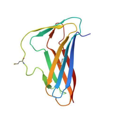
    We report the crystal structure solution of the Intracellular Protease Inhibitor (IPI) protein from Bacillus subtilis, which has been reported to be an inhibitor of the intracellular subtilisin Isp1 from the same organism. The structure of IPI is a variant of the all-beta, immunoglobulin (Ig) fold. It is possible that IPI is important for protein-protein interactions, of which inhibition of Isp1 is one. The intracellular nature of ISP is questioned, because an alternative ATG codon in the ipi gene would produce a protein with an N-terminal extension containing a signal peptide. It is possible that alternative initiation exists, producing either an intracellular inhibitor or a secreted form that may be associated with the cell surface.  Homologues of the IPI protein from other species are multi-domain proteins, containing signal peptides and domains also associated with the bacterial cell-surface. The cysteine peptidase inhibitors chagasin and amoebiasin also have Ig-like folds, but their topology differs significantly from that of IPI, and they share no recent common ancestor. A model of IPI docked to Isp1 shows similarities to other subtilisin:inhibitor complexes, particularly where the inhibitor interacts with the peptidase active site.


  • Organizational Affiliation

    Institute of Integrative Biology, University of Liverpool, Liverpool, L69 7ZB, UK.


Macromolecules
Find similar proteins by:  (by identity cutoff)  |  3D Structure
Entity ID: 1
MoleculeChains Sequence LengthOrganismDetailsImage
Intracellular proteinase inhibitor120Bacillus subtilisMutation(s): 0 
Gene Names: BSU11130ipiNP_388994.1
UniProt
Find proteins for P39804 (Bacillus subtilis (strain 168))
Explore P39804 
Go to UniProtKB:  P39804
Entity Groups  
Sequence Clusters30% Identity50% Identity70% Identity90% Identity95% Identity100% Identity
UniProt GroupP39804
Sequence Annotations
Expand
  • Reference Sequence
Small Molecules
Ligands 1 Unique
IDChains Name / Formula / InChI Key2D Diagram3D Interactions
PG4
Query on PG4

Download Ideal Coordinates CCD File 
B [auth A]TETRAETHYLENE GLYCOL
C8 H18 O5
UWHCKJMYHZGTIT-UHFFFAOYSA-N
Modified Residues  1 Unique
IDChains TypeFormula2D DiagramParent
MSE
Query on MSE
A
L-PEPTIDE LINKINGC5 H11 N O2 SeMET
Experimental Data & Validation

Experimental Data

Unit Cell:
Length ( Å )Angle ( ˚ )
a = 73.586α = 90
b = 73.586β = 90
c = 132.923γ = 90
Software Package:
Software NamePurpose
REFMACrefinement
PHENIXrefinement
SHELXphasing
MolProbitymodel building
XSCALEdata scaling
PDB_EXTRACTdata extraction
XDSdata reduction
SHELXDphasing
autoSHARPphasing

Structure Validation

View Full Validation Report


Entry History 

Deposition Data

Revision History  (Full details and data files)

  • Version 1.0: 2009-09-15
    Type: Initial release
  • Version 1.1: 2011-07-13
    Changes: Advisory, Version format compliance
  • Version 1.2: 2014-06-25
    Changes: Database references
  • Version 1.3: 2017-10-25
    Changes: Author supporting evidence, Refinement description
  • Version 1.4: 2019-07-24
    Changes: Data collection, Derived calculations, Refinement description
  • Version 1.5: 2023-02-01
    Changes: Database references, Derived calculations