Defining the role of the axial ligand of the type 1 copper site in amicyanin by replacement of methionine with leucine.
Choi, M., Sukumar, N., Liu, A., Davidson, V.L.(2009) Biochemistry 48: 9174-9184
- PubMed: 19715303
- DOI: https://doi.org/10.1021/bi900836h
- Primary Citation of Related Structures:
3IE9, 3IEA - PubMed Abstract:
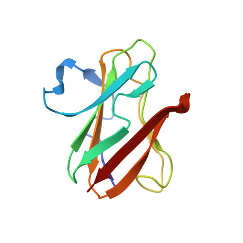
The effects of replacing the axial methionine ligand of the type 1 copper site with leucine on the structure and function of amicyanin have been characterized. The crystal structures of the oxidized and reduced forms of the protein reveal that the copper site is now tricoordinate with no axial ligand, and that the copper coordination distances for the two ligands provided by histidines are significantly increased. Despite these structural changes, the absorption and EPR spectra of M98L amicyanin are only slightly altered and still consistent with that of a typical type 1 site. The oxidation-reduction midpoint potential (E(m)) value becomes 127 mV more positive as a consequence of the M98L mutation, most likely because of the increased hydrophobicity of the copper site. The most dramatic effect of the mutation was on the electron transfer (ET) reaction from reduced M98L amicyanin to cytochrome c(551i) within the protein ET complex. The rate decreased 435-fold, which was much more than expected from the change in E(m). Examination of the temperature dependence of the ET rate (k(ET)) revealed that the mutation caused a 13.6-fold decrease in the electronic coupling (H(AB)) for the reaction. A similar decrease was predicted from a comparative analysis of the crystal structures of reduced M98L and native amicyanins. The most direct route of ET for this reaction is through the Met98 ligand. Inspection of the structures suggests that the major determinant of the large decrease in the experimentally determined values of H(AB) and k(ET) is the increased distance from the copper to the protein within the type 1 site of M98L amicyanin.
Organizational Affiliation:
Department of Biochemistry, The University of Mississippi Medical Center, Jackson, Mississippi 39216-4505, USA.


