Removing the invariant salt bridge of parvalbumin increases flexibility in the AB-loop structure
Hoh, F., Cave, A., Strub, M.P., Baneres, J.L., Padilla, A.(2009) Acta Crystallogr D Biol Crystallogr 65: 733-743
- PubMed: 19622856
- DOI: https://doi.org/10.1107/S0907444909011482
- Primary Citation of Related Structures:
3F45 - PubMed Abstract:
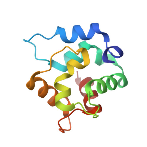
Parvalbumins (PVs) are calcium-buffer proteins that belong to the EF-hand family. Their N-terminal domain consists of two antiparallel helices A and B that make up a flat hydrophobic surface that is associated with the opposite side of the CD and EF binding sites. A single conserved Arg75-Glu81 salt bridge is buried in this hydrophobic interface. The structure of a rat PV mutant in which Arg75 was replaced by alanine was solved by molecular replacement. Unexpectedly, a large distance deviation of 7.8 A was observed for the AB loop but not for the residues that flank the R75A mutation. The thermal stability of the calcium-loaded form is lower (T(m) = 352.0 K; DeltaT(m) = -11.4 K) than that of the wild-type protein and the apo mutant is unfolded at room temperature. Weaker calcium or magnesium affinities were also measured for the R75A mutant (Ca(2+): K(1) = 4.21 x 10(7) M(-1), K(2) = 6.18 x 10(6) M(-1); Mg(2+): K(1) = 2.98 x 10(4) M(-1), K(2) = 3.09 x 10(3) M(-1)). Finally, comparison of the B factors showed an increase in the flexibility of the AB loop that is consistent with this region being more exposed to solvent in the mutant. The mutant structure therefore demonstrates the role of the salt bridge in attaching the nonbinding AB domain to the remaining protein core. Normal-mode analysis indeed indicated an altered orientation of the AB domain with regard to the CD-EF binding domains.
- CNRS UMR 5048, INSERM U554, Université Montpellier 1 et 2, Centre de Biochimie Structurale, 34090 Montpellier, France.
Organizational Affiliation: