3ETQ

X-ray structure of cysteine-free fragment of mHCN2 C-terminal region from amino acids 443-630 including C508N, C584S, and C601S mutations


Experimental Data Snapshot

  • Method: X-RAY DIFFRACTION
  • Resolution: 1.90 Å
  • R-Value Free: 0.216 
  • R-Value Work: 0.182 
  • R-Value Observed: 0.183 

wwPDB Validation   3D Report Full Report


Ligand Structure Quality Assessment 


This is version 1.4 of the entry. See complete history


Literature

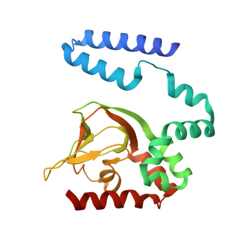
Mapping the structure and conformational movements of proteins with transition metal ion FRET.

Taraska, J.W.Puljung, M.C.Olivier, N.B.Flynn, G.E.Zagotta, W.N.

(2009) Nat Methods 6: 532-537

  • DOI: https://doi.org/10.1038/nmeth.1341
  • Primary Citation of Related Structures:  
    3ETQ, 3FFQ

  • PubMed Abstract: 

    Visualizing conformational dynamics in proteins has been difficult, and the atomic-scale motions responsible for the behavior of most allosteric proteins are unknown. Here we report that fluorescence resonance energy transfer (FRET) between a small fluorescent dye and a nickel ion bound to a dihistidine motif can be used to monitor small structural rearrangements in proteins. This method provides several key advantages over classical FRET, including the ability to measure the dynamics of close-range interactions, the use of small probes with short linkers, a low orientation dependence, and the ability to add and remove unique tunable acceptors. We used this 'transition metal ion FRET' approach along with X-ray crystallography to determine the structural changes of the gating ring of the mouse hyperpolarization-activated cyclic nucleotide-regulated ion channel HCN2. Our results suggest a general model for the conformational switch in the cyclic nucleotide-binding site of cyclic nucleotide-regulated ion channels.


  • Organizational Affiliation

    Department of Physiology and Biophysics, Howard Hughes Medical Institute, University of Washington, Seattle, Washington, USA.


Macromolecules
Find similar proteins by:  (by identity cutoff)  |  3D Structure
Entity ID: 1
MoleculeChains Sequence LengthOrganismDetailsImage
Potassium/sodium hyperpolarization-activated cyclic nucleotide-gated channel 2
A, B
204Mus musculusMutation(s): 3 
Gene Names: Bcng2Hac1Hcn2
UniProt
Find proteins for O88703 (Mus musculus)
Explore O88703 
Go to UniProtKB:  O88703
Entity Groups  
Sequence Clusters30% Identity50% Identity70% Identity90% Identity95% Identity100% Identity
UniProt GroupO88703
Sequence Annotations
Expand
  • Reference Sequence
Small Molecules
Ligands 1 Unique
IDChains Name / Formula / InChI Key2D Diagram3D Interactions
CMP
Query on CMP

Download Ideal Coordinates CCD File 
C [auth A],
D [auth B]
ADENOSINE-3',5'-CYCLIC-MONOPHOSPHATE
C10 H12 N5 O6 P
IVOMOUWHDPKRLL-KQYNXXCUSA-N
Experimental Data & Validation

Experimental Data

  • Method: X-RAY DIFFRACTION
  • Resolution: 1.90 Å
  • R-Value Free: 0.216 
  • R-Value Work: 0.182 
  • R-Value Observed: 0.183 
  • Space Group: P 4 21 2
Unit Cell:
Length ( Å )Angle ( ˚ )
a = 95.752α = 90
b = 95.752β = 90
c = 115.223γ = 90
Software Package:
Software NamePurpose
DENZOdata reduction
SCALEPACKdata scaling
PHASERphasing
REFMACrefinement
PDB_EXTRACTdata extraction
HKL-2000data scaling

Structure Validation

View Full Validation Report


Ligand Structure Quality Assessment 


Entry History 

Deposition Data

  • Released Date: 2009-06-23 
  • Deposition Author(s): Flynn, G.E.

Revision History  (Full details and data files)

  • Version 1.0: 2009-06-23
    Type: Initial release
  • Version 1.1: 2011-07-13
    Changes: Version format compliance
  • Version 1.2: 2017-10-25
    Changes: Refinement description
  • Version 1.3: 2021-10-20
    Changes: Database references, Derived calculations
  • Version 1.4: 2023-09-06
    Changes: Data collection, Refinement description