3C1V

The 1.5 A Crystal structure of Ca2+-bound S100A4


Experimental Data Snapshot

  • Method: X-RAY DIFFRACTION
  • Resolution: 1.50 Å
  • R-Value Free: 0.218 
  • R-Value Work: 0.195 
  • R-Value Observed: 0.196 

wwPDB Validation   3D Report Full Report


This is version 1.3 of the entry. See complete history


Literature

Crystal structure of the Ca(2+)-form and Ca(2+)-binding kinetics of metastasis-associated protein, S100A4

Gingras, A.R.Basran, J.Prescott, A.Kriajevska, M.Bagshaw, C.R.Barsukov, I.L.

(2008) FEBS Lett 582: 1651-1656

  • DOI: https://doi.org/10.1016/j.febslet.2008.04.017
  • Primary Citation of Related Structures:  
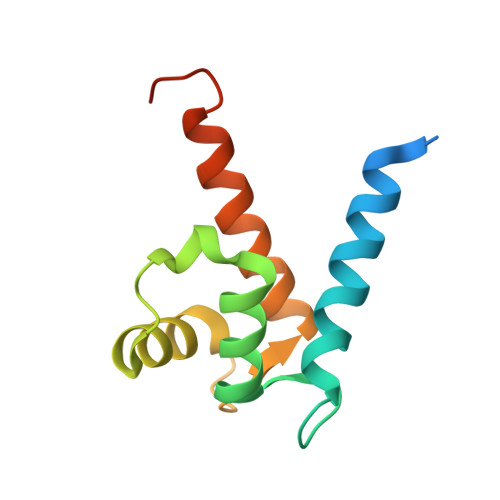
    3C1V

  • PubMed Abstract: 

    S100A4 takes part in control of tumour cell migration and contributes to metastatic spread in in vivo models. In the active dimeric Ca(2+)-bound state it interacts with multiple intracellular targets. Conversely, oligomeric forms of S100A4 are linked with the extracellular function of this protein. We report the 1.5A X-ray crystal structure of Ca(2+)-bound S100A4 and use it to identify the residues involved in target recognition and to derive a model of the oligomeric state. We applied stopped-flow analysis of tyrosine fluorescence to derive kinetics of S100A4 activation by Ca(2+) (k(on)=3.5 microM(-1)s(-1), k(off)=20s(-1)).


  • Organizational Affiliation

    Department of Biochemistry, University of Leicester, Henry Wellcome Building, Leicester LE1 9HN, UK.


Macromolecules
Find similar proteins by:  (by identity cutoff)  |  3D Structure
Entity ID: 1
MoleculeChains Sequence LengthOrganismDetailsImage
Protein S100-A4
A, B, C, D
113Homo sapiensMutation(s): 0 
Gene Names: S100A4
UniProt & NIH Common Fund Data Resources
Find proteins for P26447 (Homo sapiens)
Explore P26447 
Go to UniProtKB:  P26447
PHAROS:  P26447
GTEx:  ENSG00000196154 
Entity Groups  
Sequence Clusters30% Identity50% Identity70% Identity90% Identity95% Identity100% Identity
UniProt GroupP26447
Sequence Annotations
Expand
  • Reference Sequence
Experimental Data & Validation

Experimental Data

  • Method: X-RAY DIFFRACTION
  • Resolution: 1.50 Å
  • R-Value Free: 0.218 
  • R-Value Work: 0.195 
  • R-Value Observed: 0.196 
  • Space Group: P 32
Unit Cell:
Length ( Å )Angle ( ˚ )
a = 52.32α = 90
b = 52.32β = 90
c = 139.14γ = 120
Software Package:
Software NamePurpose
REFMACrefinement
MxCuBEdata collection
XDSdata reduction
XDSdata scaling
PHASERphasing

Structure Validation

View Full Validation Report


Entry History 

Deposition Data

Revision History  (Full details and data files)

  • Version 1.0: 2008-04-01
    Type: Initial release
  • Version 1.1: 2011-07-13
    Changes: Version format compliance
  • Version 1.2: 2018-01-31
    Changes: Experimental preparation
  • Version 1.3: 2023-11-01
    Changes: Data collection, Database references, Derived calculations, Refinement description