3B0B

Crystal structure of the chicken CENP-S/CENP-X complex


Experimental Data Snapshot

  • Method: X-RAY DIFFRACTION
  • Resolution: 2.15 Å
  • R-Value Free: 0.235 
  • R-Value Work: 0.186 
  • R-Value Observed: 0.190 

wwPDB Validation   3D Report Full Report


This is version 1.0 of the entry. See complete history


Literature

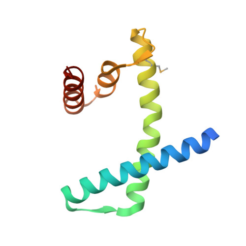
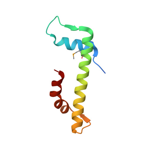
CENP-T-W-S-X Forms a Unique Centromeric Chromatin Structure with a Histone-like Fold.

Nishino, T.Takeuchi, K.Gascoigne, K.E.Suzuki, A.Hori, T.Oyama, T.Morikawa, K.Cheeseman, I.M.Fukagawa, T.

(2012) Cell 148: 487-501

  • DOI: https://doi.org/10.1016/j.cell.2011.11.061
  • Primary Citation of Related Structures:  
    3B0B, 3B0C, 3B0D, 3VH5, 3VH6

  • PubMed Abstract: 

    The multiprotein kinetochore complex must assemble at a specific site on each chromosome to achieve accurate chromosome segregation. Defining the nature of the DNA-protein interactions that specify the position of the kinetochore and provide a scaffold for kinetochore formation remain key goals. Here, we demonstrate that the centromeric histone-fold-containing CENP-T-W and CENP-S-X complexes coassemble to form a stable CENP-T-W-S-X heterotetramer. High-resolution structural analysis of the individual complexes and the heterotetramer reveals similarity to other histone fold-containing complexes including canonical histones within a nucleosome. The CENP-T-W-S-X heterotetramer binds to and supercoils DNA. Mutants designed to compromise heterotetramerization or the DNA-protein contacts around the heterotetramer strongly reduce the DNA binding and supercoiling activities in vitro and compromise kinetochore assembly in vivo. These data suggest that the CENP-T-W-S-X complex forms a unique nucleosome-like structure to generate contacts with DNA, extending the "histone code" beyond canonical nucleosome proteins.


  • Organizational Affiliation

    Department of Molecular Genetics, National Institute of Genetics and The Graduate University for Advanced Studies (SOKENDAI), Mishima, Shizuoka 411-8540, Japan.


Macromolecules
Find similar proteins by:  (by identity cutoff)  |  3D Structure
Entity ID: 1
MoleculeChains Sequence LengthOrganismDetailsImage
Centromere protein SA [auth B],
C [auth A]
107Gallus gallusMutation(s): 0 
Gene Names: CENP-S
UniProt
Find proteins for E1BSW7 (Gallus gallus)
Explore E1BSW7 
Go to UniProtKB:  E1BSW7
Entity Groups  
Sequence Clusters30% Identity50% Identity70% Identity90% Identity95% Identity100% Identity
UniProt GroupE1BSW7
Sequence Annotations
Expand
  • Reference Sequence
Find similar proteins by:  (by identity cutoff)  |  3D Structure
Entity ID: 2
MoleculeChains Sequence LengthOrganismDetailsImage
Centromere protein XB [auth C],
D
81Gallus gallusMutation(s): 0 
Gene Names: CENP-X
UniProt
Find proteins for P0DJH7 (Gallus gallus)
Explore P0DJH7 
Go to UniProtKB:  P0DJH7
Entity Groups  
Sequence Clusters30% Identity50% Identity70% Identity90% Identity95% Identity100% Identity
UniProt GroupP0DJH7
Sequence Annotations
Expand
  • Reference Sequence
Small Molecules
Modified Residues  1 Unique
IDChains TypeFormula2D DiagramParent
MSE
Query on MSE
A [auth B],
C [auth A]
L-PEPTIDE LINKINGC5 H11 N O2 SeMET
Experimental Data & Validation

Experimental Data

  • Method: X-RAY DIFFRACTION
  • Resolution: 2.15 Å
  • R-Value Free: 0.235 
  • R-Value Work: 0.186 
  • R-Value Observed: 0.190 
  • Space Group: P 61
Unit Cell:
Length ( Å )Angle ( ˚ )
a = 48.741α = 90
b = 48.741β = 90
c = 345.633γ = 120
Software Package:
Software NamePurpose
HKL-2000data collection
SOLVEphasing
PHENIXrefinement
HKL-2000data reduction
HKL-2000data scaling

Structure Validation

View Full Validation Report


Entry History 

Deposition Data

Revision History  (Full details and data files)

  • Version 1.0: 2012-03-07
    Type: Initial release