Download Mendeley
Structural and functional insights into a mesophilic cold shock protein CspA with enhanced precision.
Wanko Nembot, M., Feller, G., Volkov, A.N., le Paige, U.B., Bouvignies, G., Damblon, C.(2026) J Magn Reson (1969 388: 108077-108077
- PubMed: 42102648 Search on PubMed
- DOI: https://doi.org/10.1016/j.jmr.2026.108077
- Primary Citation Related Structures:
30IB - PubMed Abstract:
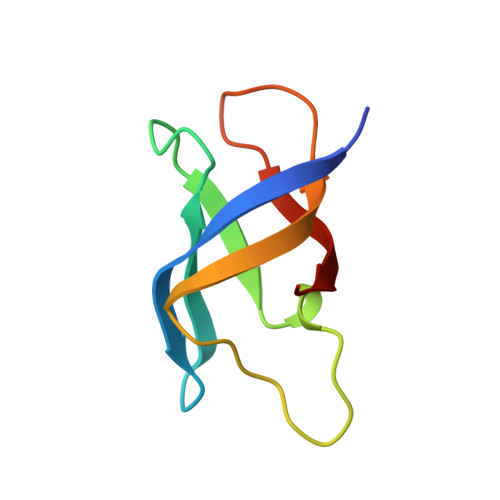
Cold-shock proteins (CSPs) are highly conserved nucleic acid-binding proteins that act as chaperones during cellular adaptation to low temperatures. Here, we present a comprehensive structural and dynamic characterization of Escherichia coli CspA using high-resolution NMR spectroscopy. The solution structure of CspA (PDB ID: 30IB) is supported by extensive NMR experimental restraints, minimal violations, and favorable stereochemistry, establishing it as a well-converged NMR structure. Crucially, we investigated backbone dynamics across multiple timescales, with a particular focus on the microsecond-millisecond regime using a combination of 15 N Carr-Purcell-Meiboom-Gill (CPMG) relaxation dispersion and 15 N chemical exchange saturation transfer (CEST) experiments, together with visible peak-position constraints. To our knowledge, this represents the first application of such a combined 15 N CEST, 15 N CPMG and visible peak-position constraints approach to probe conformational exchange in CSPs. Our results show that, in addition to the conserved aromatic residues of RNP1 and RNP2 motifs that mediate π-stacking interactions with nucleic acids, an unexpectedly broad network of hydrophobic core and solvent-exposed polar residues undergoes conformational exchange. Notably, residues in the β3-β4 and β4-β5 loops display complex dynamics not fully captured by model-free analysis formalism, highlighting their role in binding site flexibility. Complementary AF3/YASARA modeling of the CspA bound to heptathymidine (dT7) further supported that aromatic and polar residues form π-stacking and ionic interactions with ssDNA bases, corroborating the functional relevance of these dynamic regions. Therefore, our findings demonstrate that CspA relies on a dynamic network extending from conserved motifs through the hydrophobic core and flexible loops, conferring the structural adaptability required for efficient nucleic acid recognition and chaperone activity.
- Structural Biological Chemistry Laboratory, MolSys Research Unity, University of Liege, Liège, Belgium. Electronic address: amwanko@uliege.be.
Organizational Affiliation: