


Crystal structure of D-alanine:D-Alanine Ligase with AMPPNP from Thermus thermophilus HB8
Kitamura, Y., Yokoyama, S., Kuramitsu, S.To be published.
Experimental Data Snapshot
Entity ID: 1 | |||||
|---|---|---|---|---|---|
| Molecule | Chains | Sequence Length | Organism | Details | Image |
| D-alanine-D-alanine ligase | 319 | Thermus thermophilus HB8 | Mutation(s): 0 EC: 6.3.2.4 | 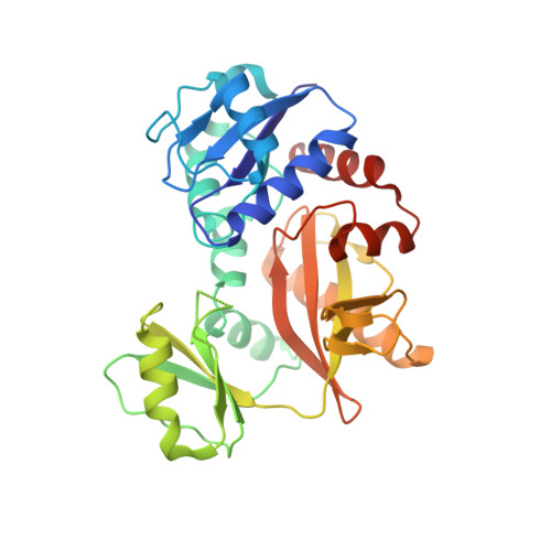 | |
UniProt | |||||
Find proteins for Q5SHZ3 (Thermus thermophilus (strain ATCC 27634 / DSM 579 / HB8)) Explore Q5SHZ3 Go to UniProtKB: Q5SHZ3 | |||||
Entity Groups | |||||
| Sequence Clusters | 30% Identity50% Identity70% Identity90% Identity95% Identity100% Identity | ||||
| UniProt Group | Q5SHZ3 | ||||
Sequence AnnotationsExpand | |||||
| |||||
| Ligands 1 Unique | |||||
|---|---|---|---|---|---|
| ID | Chains | Name / Formula / InChI Key | 2D Diagram | 3D Interactions | |
| ANP Query on ANP | D [auth A], E [auth B], F [auth C] | PHOSPHOAMINOPHOSPHONIC ACID-ADENYLATE ESTER C10 H17 N6 O12 P3 PVKSNHVPLWYQGJ-KQYNXXCUSA-N |  | ||
| Length ( Å ) | Angle ( ˚ ) |
|---|---|
| a = 167.72 | α = 90 |
| b = 56.708 | β = 108.56 |
| c = 141.826 | γ = 90 |
| Software Name | Purpose |
|---|---|
| CNS | refinement |
| HKL-2000 | data collection |
| HKL-2000 | data reduction |
| HKL-2000 | data scaling |
| MOLREP | phasing |
RCSB PDB (citation) is hosted by
RCSB PDB is a member of the

