Conservation of Dark Recovery Kinetic Parameters and Structural Features in the Pseudomonadaceae "Short" Light, Oxygen, Voltage (Lov) Protein Family: Implications for the Design of Lov-Based Optogenetic Tools.
Rani, R., Jentzsch, K., Lecher, J., Hartmann, R., Willbold, D., Jaeger, K., Krauss, U.(2013) Biochemistry 52: 4460
- PubMed: 23746326
- DOI: https://doi.org/10.1021/bi400311r
- Primary Citation of Related Structures:
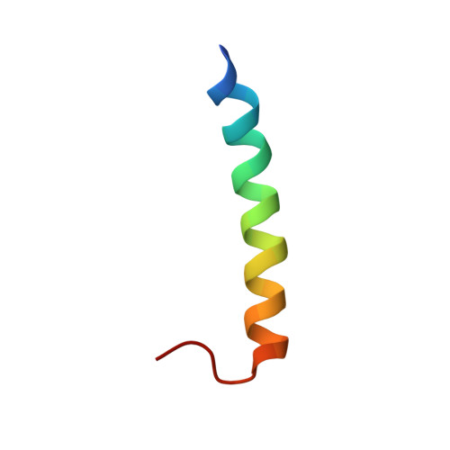
2YOM, 2YON - PubMed Abstract:
In bacteria and fungi, various light, oxygen, voltage (LOV) sensory systems that lack a fused effector domain but instead contain only short N- and C-terminal extensions flanking the LOV core exist. In the prokaryotic kingdom, this so-called "short" LOV protein family represents the third largest LOV photoreceptor family. This observation prompted us to study their distribution and phylogeny as well as their photochemical and structural properties in more detail. We recently described the slow and fast reverting "short" LOV proteins PpSB1-LOV and PpSB2-LOV from Pseudomonas putida KT2440 whose adduct state lifetimes varied by 3 orders of magnitude [Jentzsch, K., Wirtz, A., Circolone, F., Drepper, T., Losi, A., Gärtner, W., Jaeger, K. E., and Krauss, U. (2009) Biochemistry 48, 10321-10333]. We now present evidence of the conservation of similar fast and slow-reverting "short" LOV proteins in different Pseudomonas species. Truncation studies conducted with PpSB1-LOV and PpSB2-LOV suggested that the short N- and C-terminal extensions outside of the LOV core domain are essential for the structural integrity and folding of the two proteins. While circular dichroism and solution nuclear magnetic resonance experiments verify that the two short C-terminal extensions of PpSB1-LOV and PpSB2-LOV form independently folding helical structures in solution, bioinformatic analyses imply the formation of coiled coils of the respective structural elements in the context of the dimeric full-length proteins. Given their prototypic architecture, conserved in most more complex LOV photoreceptor systems, "short" LOV proteins could represent ideally suited building blocks for the design of genetically encoded photoswitches (i.e., LOV-based optogenetic tools).
- Institut für Molekulare Enzymtechnologie, Heinrich-Heine-Universität Düsseldorf, Forschungszentrum Jülich, Stetternicher Forst, D-52426 Jülich, Germany.
Organizational Affiliation: