2YL2

Crystal structure of human acetyl-CoA carboxylase 1, biotin carboxylase (BC) domain


Experimental Data Snapshot

  • Method: X-RAY DIFFRACTION
  • Resolution: 2.30 Å
  • R-Value Free: 0.241 
  • R-Value Work: 0.206 
  • R-Value Observed: 0.208 

wwPDB Validation   3D Report Full Report


This is version 1.2 of the entry. See complete history


Literature

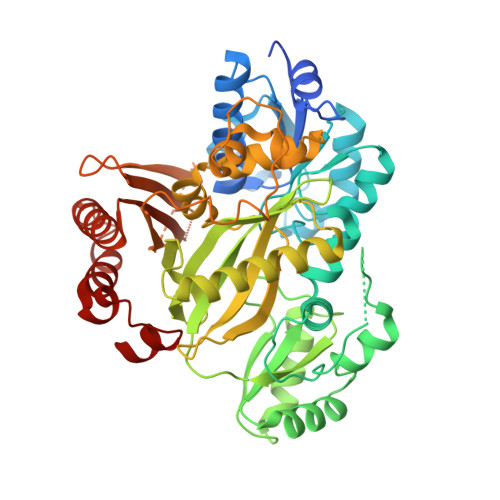
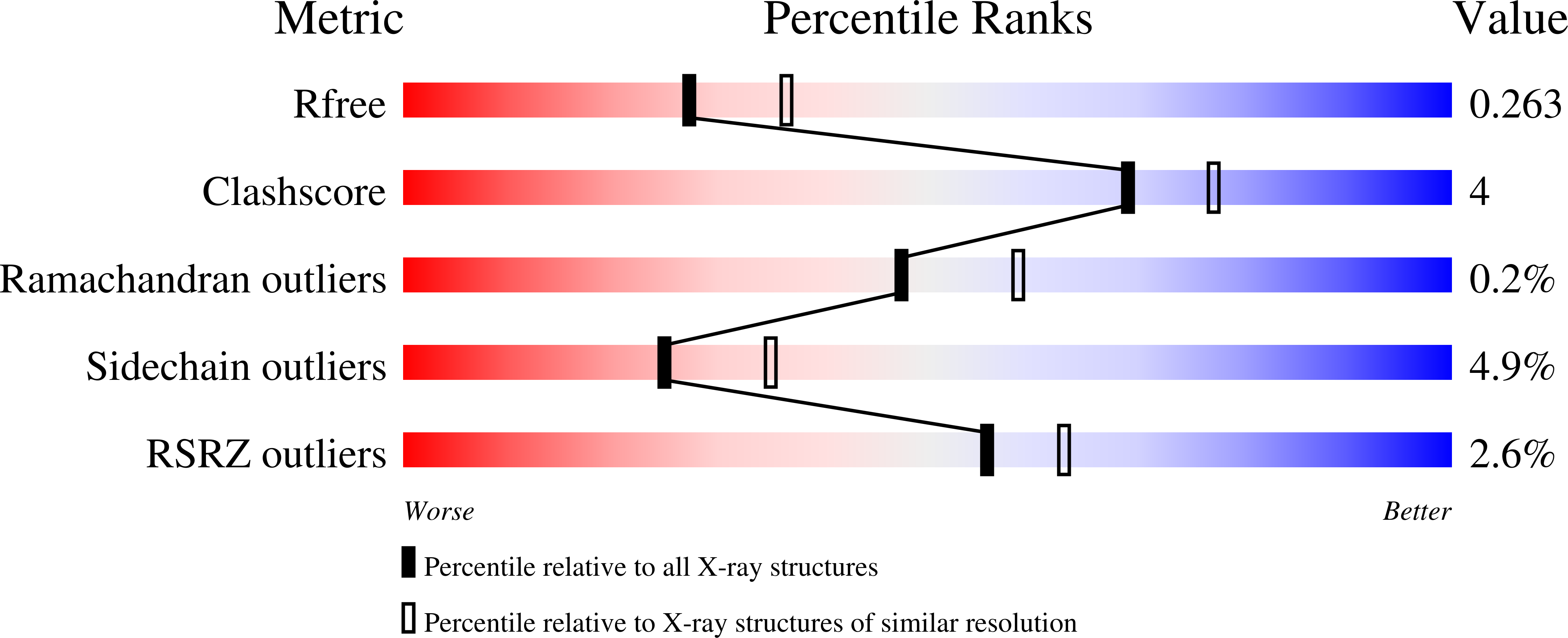
Crystal Structure of Human Acetyl-Coa Carboxylase 1, Biotin Carboxylase (Bc) Domain

Muniz, J.R.C.Froese, D.S.Krysztofinska, E.Vollmar, M.Beltrami, A.Krojer, T.Allerston, C.K.von Delft, F.Arrowsmith, C.H.Edwards, A.M.Weigelt, J.Bountra, C.Yue, W.W.Oppermann, U.

To be published.

Macromolecules
Find similar proteins by:  (by identity cutoff)  |  3D Structure
Entity ID: 1
MoleculeChains Sequence LengthOrganismDetailsImage
ACETYL-COA CARBOXYLASE 1
A, B
540Homo sapiensMutation(s): 0 
EC: 6.4.1.2 (PDB Primary Data), 6.3.4.14 (PDB Primary Data)
UniProt & NIH Common Fund Data Resources
Find proteins for Q13085 (Homo sapiens)
Explore Q13085 
Go to UniProtKB:  Q13085
PHAROS:  Q13085
GTEx:  ENSG00000278540 
Entity Groups  
Sequence Clusters30% Identity50% Identity70% Identity90% Identity95% Identity100% Identity
UniProt GroupQ13085
Sequence Annotations
Expand
  • Reference Sequence
Experimental Data & Validation

Experimental Data

  • Method: X-RAY DIFFRACTION
  • Resolution: 2.30 Å
  • R-Value Free: 0.241 
  • R-Value Work: 0.206 
  • R-Value Observed: 0.208 
  • Space Group: P 1
Unit Cell:
Length ( Å )Angle ( ˚ )
a = 60.67α = 92.04
b = 60.87β = 98
c = 70.37γ = 92.2
Software Package:
Software NamePurpose
BUSTERrefinement
MOSFLMdata reduction
SCALAdata scaling
PHASERphasing

Structure Validation

View Full Validation Report


Entry History 

Revision History  (Full details and data files)

  • Version 1.0: 2011-06-15
    Type: Initial release
  • Version 1.1: 2011-07-13
    Changes: Version format compliance
  • Version 1.2: 2018-01-24
    Changes: Database references