2XZK

THE ASPERGILLUS FUMIGATUS SIALIDASE IS A KDNASE: STRUCTURAL AND MECHANISTIC INSIGHTS


Experimental Data Snapshot

  • Method: X-RAY DIFFRACTION
  • Resolution: 1.50 Å
  • R-Value Free: 0.214 
  • R-Value Work: 0.179 
  • R-Value Observed: 0.181 

wwPDB Validation   3D Report Full Report


Ligand Structure Quality Assessment 


This is version 1.4 of the entry. See complete history


Literature

The Aspergillus Fumigatus Sialidase is a Kdnase: Structural and Mechanistic Insights.

Telford, J.C.Yeung, J.H.F.Xu, G.Kiefel, M.J.Watts, A.G.Hader, S.Chan, J.Bennet, A.J.Moore, M.M.Taylor, G.L.

(2011) J Biol Chem 286: 10783

  • DOI: https://doi.org/10.1074/jbc.M110.207043
  • Primary Citation of Related Structures:  
    2XCY, 2XZI, 2XZJ, 2XZK

  • PubMed Abstract: 

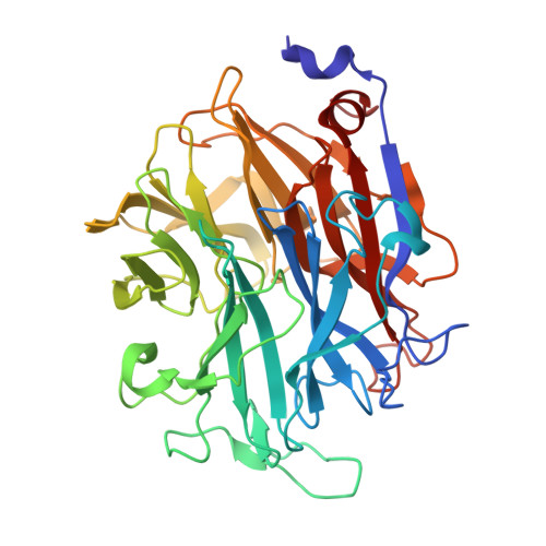
    Aspergillus fumigatus is a filamentous fungus that can cause severe respiratory disease in immunocompromised individuals. A putative sialidase from A. fumigatus was recently cloned and shown to be relatively poor in cleaving N-acetylneuraminic acid (Neu5Ac) in comparison with bacterial sialidases. Here we present the first crystal structure of a fungal sialidase. When the apo structure was compared with bacterial sialidase structures, the active site of the Aspergillus enzyme suggested that Neu5Ac would be a poor substrate because of a smaller pocket that normally accommodates the acetamido group of Neu5Ac in sialidases. A sialic acid with a hydroxyl in place of an acetamido group is 2-keto-3-deoxynononic acid (KDN). We show that KDN is the preferred substrate for the A. fumigatus sialidase and that A. fumigatus can utilize KDN as a sole carbon source. A 1.45-Å resolution crystal structure of the enzyme in complex with KDN reveals KDN in the active site in a boat conformation and nearby a second binding site occupied by KDN in a chair conformation, suggesting that polyKDN may be a natural substrate. The enzyme is not inhibited by the sialidase transition state analog 2-deoxy-2,3-dehydro-N-acetylneuraminic acid (Neu5Ac2en) but is inhibited by the related 2,3-didehydro-2,3-dideoxy-D-glycero-D-galacto-nonulosonic acid that we show bound to the enzyme in a 1.84-Å resolution crystal structure. Using a fluorinated KDN substrate, we present a 1.5-Å resolution structure of a covalently bound catalytic intermediate. The A. fumigatus sialidase is therefore a KDNase with a similar catalytic mechanism to Neu5Ac exosialidases, and this study represents the first structure of a KDNase.


  • Organizational Affiliation

    Biomedical Sciences Research Complex, University of St. Andrews, St. Andrews, Fife KY16 9ST, United Kingdom.


Macromolecules
Find similar proteins by:  (by identity cutoff)  |  3D Structure
Entity ID: 1
MoleculeChains Sequence LengthOrganismDetailsImage
EXTRACELLULAR SIALIDASE/NEURAMINIDASE, PUTATIVE
A, B
386Aspergillus fumigatus Af293Mutation(s): 0 
EC: 3.2.1.18
UniProt
Find proteins for Q4WQS0 (Aspergillus fumigatus (strain ATCC MYA-4609 / CBS 101355 / FGSC A1100 / Af293))
Explore Q4WQS0 
Go to UniProtKB:  Q4WQS0
Entity Groups  
Sequence Clusters30% Identity50% Identity70% Identity90% Identity95% Identity100% Identity
UniProt GroupQ4WQS0
Sequence Annotations
Expand
  • Reference Sequence
Small Molecules
Ligands 6 Unique
IDChains Name / Formula / InChI Key2D Diagram3D Interactions
K99
Query on K99

Download Ideal Coordinates CCD File 
D [auth A],
E [auth A],
K [auth B],
L [auth B],
S [auth B]
(2R,3R,4R,5R,6S)-2,3-bis(fluoranyl)-4,5-bis(oxidanyl)-6-[(1R,2R)-1,2,3-tris(oxidanyl)propyl]oxane-2-carboxylic acid
C9 H14 F2 O8
KSUIKNBQJWNZMW-AWLRDIFTSA-N
FKD
Query on FKD

Download Ideal Coordinates CCD File 
C [auth A],
J [auth B]
3-deoxy-3-fluoro-D-erythro-alpha-L-manno-non-2-ulopyranosonic acid
C9 H15 F O9
KOWJBKIDVGQXJZ-QMFVTVPYSA-N
GOL
Query on GOL

Download Ideal Coordinates CCD File 
F [auth A],
M [auth B],
N [auth B],
O [auth B],
P [auth B]
GLYCEROL
C3 H8 O3
PEDCQBHIVMGVHV-UHFFFAOYSA-N
NO3
Query on NO3

Download Ideal Coordinates CCD File 
G [auth A]NITRATE ION
N O3
NHNBFGGVMKEFGY-UHFFFAOYSA-N
CL
Query on CL

Download Ideal Coordinates CCD File 
I [auth A],
R [auth B]
CHLORIDE ION
Cl
VEXZGXHMUGYJMC-UHFFFAOYSA-M
NA
Query on NA

Download Ideal Coordinates CCD File 
H [auth A],
Q [auth B]
SODIUM ION
Na
FKNQFGJONOIPTF-UHFFFAOYSA-N
Experimental Data & Validation

Experimental Data

  • Method: X-RAY DIFFRACTION
  • Resolution: 1.50 Å
  • R-Value Free: 0.214 
  • R-Value Work: 0.179 
  • R-Value Observed: 0.181 
  • Space Group: P 1 21 1
Unit Cell:
Length ( Å )Angle ( ˚ )
a = 75.68α = 90
b = 58.08β = 100.05
c = 94.45γ = 90
Software Package:
Software NamePurpose
REFMACrefinement
MOSFLMdata reduction
SCALAdata scaling
REFMACphasing

Structure Validation

View Full Validation Report


Ligand Structure Quality Assessment 


Entry History 

Deposition Data

Revision History  (Full details and data files)

  • Version 1.0: 2011-01-19
    Type: Initial release
  • Version 1.1: 2011-08-17
    Changes: Database references, Non-polymer description, Version format compliance
  • Version 1.2: 2012-05-16
    Changes: Other
  • Version 1.3: 2020-07-29
    Type: Remediation
    Reason: Carbohydrate remediation
    Changes: Derived calculations, Other, Structure summary
  • Version 1.4: 2023-12-20
    Changes: Data collection, Database references, Refinement description, Structure summary