2XQW

Structure of Factor H domains 19-20 in complex with complement C3d


Experimental Data Snapshot

  • Method: X-RAY DIFFRACTION
  • Resolution: 2.31 Å
  • R-Value Free: 0.242 
  • R-Value Work: 0.201 
  • R-Value Observed: 0.203 

wwPDB Validation   3D Report Full Report


This is version 1.3 of the entry. See complete history


Literature

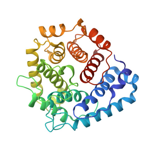
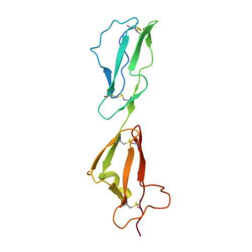
Dual Interaction of Factor H with C3D and Glycosaminoglycans in Host-Nonhost Discrimination by Complement.

Kajander, T.Lehtinen, M.J.Hyvarinen, S.Bhattacharjee, A.Leung, E.Isenman, D.E.Meri, S.Goldman, A.Jokiranta, T.S.

(2011) Proc Natl Acad Sci U S A 108: 2897

  • DOI: https://doi.org/10.1073/pnas.1017087108
  • Primary Citation of Related Structures:  
    2XQW

  • PubMed Abstract: 

    The alternative pathway of complement is important in innate immunity, attacking not only microbes but all unprotected biological surfaces through powerful amplification. It is unresolved how host and nonhost surfaces are distinguished at the molecular level, but key components are domains 19-20 of the complement regulator factor H (FH), which interact with host (i.e., nonactivator surface glycosaminoglycans or sialic acids) and the C3d part of C3b. Our structure of the FH19-20:C3d complex at 2.3-Å resolution shows that FH19-20 has two distinct binding sites, FH19 and FH20, for C3b. We show simultaneous binding of FH19 to C3b and FH20 to nonactivator surface glycosaminoglycans, and we show that both of these interactions are necessary for full binding of FH to C3b on nonactivator surfaces (i.e., for target discrimination). We also show that C3d could replace glycosaminoglycan binding to FH20, thus providing a feedback control for preventing excess C3b deposition and complement amplification. This explains the molecular basis of atypical hemolytic uremic syndrome, where mutations on the binding interfaces between FH19-20 and C3d or between FH20 and glycosaminoglycans lead to complement attack against host surfaces.


  • Organizational Affiliation

    Institute of Biotechnology, University of Helsinki, Viikinkaari, FIN-00014, Helsinki, Finland.


Macromolecules
Find similar proteins by:  (by identity cutoff)  |  3D Structure
Entity ID: 1
MoleculeChains Sequence LengthOrganismDetailsImage
COMPLEMENT C3
A, B
294Homo sapiensMutation(s): 1 
UniProt & NIH Common Fund Data Resources
Find proteins for P01024 (Homo sapiens)
Explore P01024 
Go to UniProtKB:  P01024
PHAROS:  P01024
GTEx:  ENSG00000125730 
Entity Groups  
Sequence Clusters30% Identity50% Identity70% Identity90% Identity95% Identity100% Identity
UniProt GroupP01024
Sequence Annotations
Expand
  • Reference Sequence
Find similar proteins by:  (by identity cutoff)  |  3D Structure
Entity ID: 2
MoleculeChains Sequence LengthOrganismDetailsImage
COMPLEMENT FACTOR H133Homo sapiensMutation(s): 2 
UniProt & NIH Common Fund Data Resources
Find proteins for P08603 (Homo sapiens)
Explore P08603 
Go to UniProtKB:  P08603
PHAROS:  P08603
GTEx:  ENSG00000000971 
Entity Groups  
Sequence Clusters30% Identity50% Identity70% Identity90% Identity95% Identity100% Identity
UniProt GroupP08603
Sequence Annotations
Expand
  • Reference Sequence
Experimental Data & Validation

Experimental Data

  • Method: X-RAY DIFFRACTION
  • Resolution: 2.31 Å
  • R-Value Free: 0.242 
  • R-Value Work: 0.201 
  • R-Value Observed: 0.203 
  • Space Group: P 61
Unit Cell:
Length ( Å )Angle ( ˚ )
a = 103.554α = 90
b = 103.554β = 90
c = 141.211γ = 120
Software Package:
Software NamePurpose
PHENIXrefinement
XDSdata reduction
XDSdata scaling
PHASERphasing

Structure Validation

View Full Validation Report


Entry History 

Deposition Data

Revision History  (Full details and data files)

  • Version 1.0: 2011-02-02
    Type: Initial release
  • Version 1.1: 2011-08-17
    Changes: Database references, Version format compliance
  • Version 1.2: 2019-05-08
    Changes: Data collection, Experimental preparation, Other
  • Version 1.3: 2023-12-20
    Changes: Data collection, Database references, Other, Refinement description