The Amino-Terminal Disease Hotspot of Ryanodine Receptors Forms a Cytoplasmic Vestibule.
Tung, C.C., Lobo, P.A., Kimlicka, L., Van Petegem, F.(2010) Nature 468: 585
- PubMed: 21048710
- DOI: https://doi.org/10.1038/nature09471
- Primary Citation of Related Structures:
2XOA - PubMed Abstract:
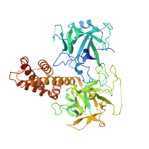
Many physiological events require transient increases in cytosolic Ca(2+) concentrations. Ryanodine receptors (RyRs) are ion channels that govern the release of Ca(2+) from the endoplasmic and sarcoplasmic reticulum. Mutations in RyRs can lead to severe genetic conditions that affect both cardiac and skeletal muscle, but locating the mutated residues in the full-length channel structure has been difficult. Here we show the 2.5 Å resolution crystal structure of a region spanning three domains of RyR type 1 (RyR1), encompassing amino acid residues 1-559. The domains interact with each other through a predominantly hydrophilic interface. Docking in RyR1 electron microscopy maps unambiguously places the domains in the cytoplasmic portion of the channel, forming a 240-kDa cytoplasmic vestibule around the four-fold symmetry axis. We pinpoint the exact locations of more than 50 disease-associated mutations in full-length RyR1 and RyR2. The mutations can be classified into three groups: those that destabilize the interfaces between the three amino-terminal domains, disturb the folding of individual domains or affect one of six interfaces with other parts of the receptor. We propose a model whereby the opening of a RyR coincides with allosterically coupled motions within the N-terminal domains. This process can be affected by mutations that target various interfaces within and across subunits. The crystal structure provides a framework to understand the many disease-associated mutations in RyRs that have been studied using functional methods, and will be useful for developing new strategies to modulate RyR function in disease states.
Organizational Affiliation:
Department of Biochemistry and Molecular Biology, University of British Columbia, Vancouver, British Columbia V6T 1Z3, Canada.