2X9D

Tet repressor (class D) in complex with iso-7-chlortetracycline


Experimental Data Snapshot

  • Method: X-RAY DIFFRACTION
  • Resolution: 2.34 Å
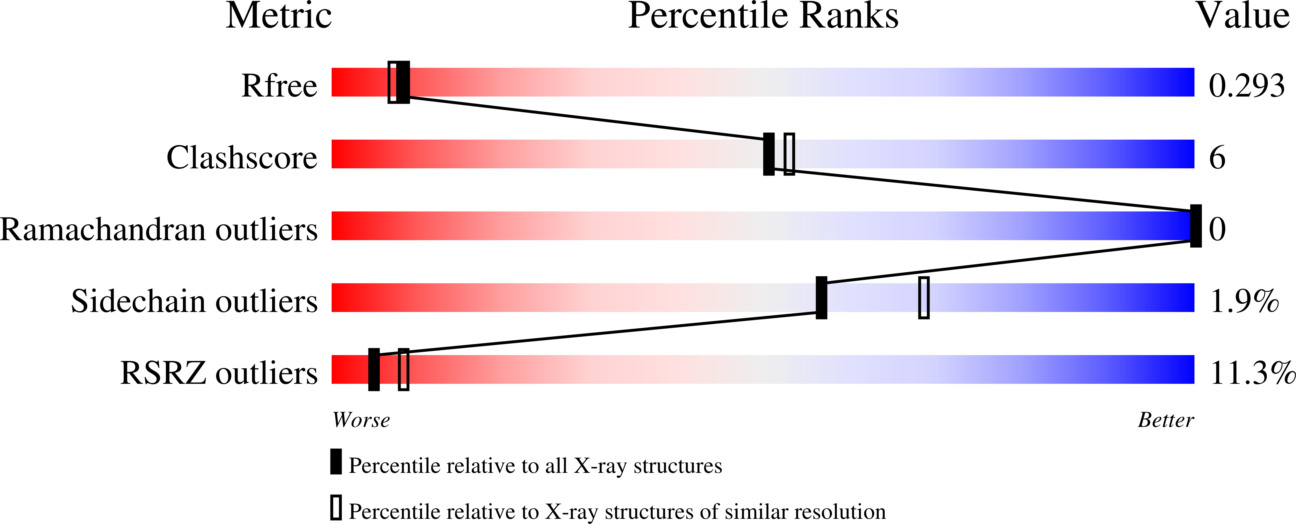
  • R-Value Free: 0.280 
  • R-Value Work: 0.234 
  • R-Value Observed: 0.238 

wwPDB Validation   3D Report Full Report


Ligand Structure Quality Assessment 


This is version 1.4 of the entry. See complete history


Literature

Recognition of Drug Degradation Products by Target Proteins: Isotetracycline Binding to Tet Repressor.

Volkers, G.Petruschka, L.Hinrichs, W.

(2011) J Med Chem 54: 5108

  • DOI: https://doi.org/10.1021/jm200332e
  • Primary Citation of Related Structures:  
    2X6O, 2X9D

  • PubMed Abstract: 

    Tetracycline antibiotics and their degradation products appear in medically treated tissues, food, soil, and manure sludge in the environment. In the context of protein interactions with various tetracyclines we performed crystal structure analyses of the tetracycline repressor in complex with weak or noninducing tetracycline derivatives. Isotetracyclines are degradation products of tetracyclines, which occur under physiological conditions. The typical framework of the antibiotic is irreversibly broken at the BC-ring connection, leading to a modified orientation of the AB to the new C*D ring fragments. The shape of the zwitterionic AB-ring fragment is unchanged and still binds to the TetR recognition site in a manner comparable to the intact antibiotic but without typical Mg(2+) chelation. This work is an example that drug degradation products can still bind to specific targets and should be discussed in light of potential and critical side effects.


  • Organizational Affiliation

    Department of Molecular Structural Biology, Institute for Biochemistry, University of Greifswald, Felix-Hausdorff-Strasse 4, D-17489 Greifswald, Germany.


Macromolecules
Find similar proteins by:  (by identity cutoff)  |  3D Structure
Entity ID: 1
MoleculeChains Sequence LengthOrganismDetailsImage
TETRACYCLINE REPRESSOR PROTEIN CLASS D207Escherichia coliMutation(s): 1 
UniProt
Find proteins for P0ACT4 (Escherichia coli)
Explore P0ACT4 
Go to UniProtKB:  P0ACT4
Entity Groups  
Sequence Clusters30% Identity50% Identity70% Identity90% Identity95% Identity100% Identity
UniProt GroupP0ACT4
Sequence Annotations
Expand
  • Reference Sequence
Experimental Data & Validation

Experimental Data

  • Method: X-RAY DIFFRACTION
  • Resolution: 2.34 Å
  • R-Value Free: 0.280 
  • R-Value Work: 0.234 
  • R-Value Observed: 0.238 
  • Space Group: I 41 2 2
Unit Cell:
Length ( Å )Angle ( ˚ )
a = 68.253α = 90
b = 68.253β = 90
c = 179.271γ = 90
Software Package:
Software NamePurpose
REFMACrefinement
DENZOdata reduction
SCALEPACKdata scaling
REFMACphasing

Structure Validation

View Full Validation Report


Ligand Structure Quality Assessment 


Entry History 

Deposition Data

Revision History  (Full details and data files)

  • Version 1.0: 2011-03-23
    Type: Initial release
  • Version 1.1: 2012-02-01
    Changes: Database references, Version format compliance
  • Version 1.2: 2019-05-08
    Changes: Data collection, Experimental preparation, Other
  • Version 1.3: 2019-07-24
    Changes: Data collection
  • Version 1.4: 2023-12-20
    Changes: Data collection, Database references, Derived calculations, Other, Refinement description