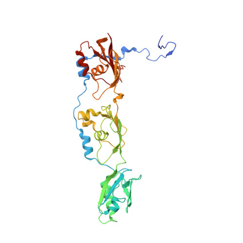
2W8H

Crystal structure of spin labeled Wza24-345.


Experimental Data Snapshot

  • Method: X-RAY DIFFRACTION
  • Resolution: 2.76 Å
  • R-Value Free: 0.229 
  • R-Value Work: 0.211 
  • R-Value Observed: 0.212 

wwPDB Validation 


Ligand Structure Quality Assessment 


This is version 1.3 of the entry. See complete history


Literature

Peldor Distance Fingerprinting of the Octameric Outer-Membrane Protein Wza from Escherichia Coli.

Hagelueken, G.Ingledew, W.J.Huang, H.Petrovic-Stojanovska, B.Whitfield, C.Elmkami, H.Schiemann, O.Naismith, J.H.

(2009) Angew Chem Int Ed Engl 48: 2904

  • DOI: https://doi.org/10.1002/anie.200805758
  • Primary Citation of Related Structures:  
    2W8H, 2W8I

  • PubMed Abstract: 

    Distance fingerprinting: Pulsed electron-electron double resonance spectroscopy (PELDOR) is applied to the octameric membrane protein complex Wza of E. coli. The data yielded a detailed distance fingerprint of its periplasmic region that compares favorably to the crystal structure. These results provide the foundation to study conformation changes from interaction with partner proteins.


  • Organizational Affiliation

    Centre for Biomolecular Sciences, The University of St. Andrews, Fife KY16 9RH, UK.


Macromolecules
Find similar proteins by:  (by identity cutoff)  |  3D Structure
Entity ID: 1
MoleculeChains Sequence LengthOrganismDetailsImage
PUTATIVE OUTER MEMBRANE LIPOPROTEIN WZA
A, B, C, D, E
A, B, C, D, E, F, G, H
329Escherichia coliMutation(s): 1 
Membrane Entity: Yes 
UniProt
Find proteins for Q9X4B7 (Escherichia coli)
Explore Q9X4B7 
Go to UniProtKB:  Q9X4B7
Entity Groups  
Sequence Clusters30% Identity50% Identity70% Identity90% Identity95% Identity100% Identity
UniProt GroupQ9X4B7
Sequence Annotations
Expand
  • Reference Sequence
Small Molecules
Ligands 2 Unique
IDChains Name / Formula / InChI Key2D Diagram3D Interactions
MTN
Query on MTN

Download Ideal Coordinates CCD File 
I [auth A]
J [auth B]
K [auth C]
L [auth D]
N [auth E]
I [auth A],
J [auth B],
K [auth C],
L [auth D],
N [auth E],
O [auth F],
P [auth G],
Q [auth H]
S-[(1-oxyl-2,2,5,5-tetramethyl-2,5-dihydro-1H-pyrrol-3-yl)methyl] methanesulfonothioate
C10 H18 N O3 S2
MXZPGYFBZHBAQM-UHFFFAOYSA-N
CL
Query on CL

Download Ideal Coordinates CCD File 
M [auth D]CHLORIDE ION
Cl
VEXZGXHMUGYJMC-UHFFFAOYSA-M
Experimental Data & Validation

Experimental Data

  • Method: X-RAY DIFFRACTION
  • Resolution: 2.76 Å
  • R-Value Free: 0.229 
  • R-Value Work: 0.211 
  • R-Value Observed: 0.212 
  • Space Group: P 21 21 2
Unit Cell:
Length ( Å )Angle ( ˚ )
a = 141.153α = 90
b = 141.152β = 90
c = 167.529γ = 90
Software Package:
Software NamePurpose
PHENIXrefinement
HKL-2000data reduction
HKL-2000data scaling
PHASERphasing

Structure Validation


Ligand Structure Quality Assessment 


Entry History 

Deposition Data

Revision History  (Full details and data files)

  • Version 1.0: 2009-02-10
    Type: Initial release
  • Version 1.1: 2011-05-12
    Changes: Version format compliance
  • Version 1.2: 2011-07-13
    Changes: Version format compliance
  • Version 1.3: 2023-12-13
    Changes: Advisory, Data collection, Database references, Derived calculations, Other, Refinement description