2W51

Human mesencephalic astrocyte-derived neurotrophic factor (MANF)


Experimental Data Snapshot

  • Method: X-RAY DIFFRACTION
  • Resolution: 2.80 Å
  • R-Value Free: 0.305 
  • R-Value Work: 0.280 
  • R-Value Observed: 0.281 

wwPDB Validation   3D Report Full Report


This is version 1.4 of the entry. See complete history


Literature

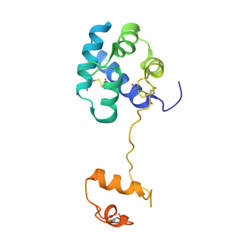
The Structure of the Conserved Neurotrophic Factors Manf and Cdnf Explains Why They are Bifunctional.

Parkash, V.Lindholm, P.Peranen, J.Kalkkinen, N.Oksanen, E.Saarma, M.Leppanen, V.M.Goldman, A.

(2009) Protein Eng Des Sel 22: 233-241

  • DOI: https://doi.org/10.1093/protein/gzn080
  • Primary Citation of Related Structures:  
    2W50, 2W51

  • PubMed Abstract: 

    We have solved the structures of mammalian mesencephalic astrocyte-derived neurotrophic factor (MANF) and conserved dopamine neurotrophic factor (CDNF). CDNF protects and repairs midbrain dopaminergic neurons in vivo; MANF supports their survival in culture and is also cytoprotective against endoplasmic reticulum (ER) stress. Neither protein structure resembles any known growth factor but the N-terminal domain is a saposin-like lipid-binding domain. MANF and CDNF may thus bind lipids or membranes. Consistent with this, there are two patches of conserved lysines and arginines. The natively unfolded MANF C-terminus contains a CKGC disulphide bridge, such as reductases and disulphide isomerases, consistent with a role in ER stress response. The structure thus explains why MANF and CDNF are bifunctional; neurotrophic activity may reside in the N-terminal domain and ER stress response in the C-terminal domain. Finally, we identified three changes, (MANF)I10-->K(CDNF), (MANF)E79-->M(CDNF) and (MANF)K88-->L(CDNF), that may account for the biological differences between the proteins.


  • Organizational Affiliation

    Institute of Biotechnology, University of Helsinki, Helsinki, Finland.


Macromolecules
Find similar proteins by:  (by identity cutoff)  |  3D Structure
Entity ID: 1
MoleculeChains Sequence LengthOrganismDetailsImage
PROTEIN ARMET158Homo sapiensMutation(s): 0 
UniProt & NIH Common Fund Data Resources
Find proteins for P55145 (Homo sapiens)
Explore P55145 
Go to UniProtKB:  P55145
PHAROS:  P55145
GTEx:  ENSG00000145050 
Entity Groups  
Sequence Clusters30% Identity50% Identity70% Identity90% Identity95% Identity100% Identity
UniProt GroupP55145
Sequence Annotations
Expand
  • Reference Sequence
Experimental Data & Validation

Experimental Data

  • Method: X-RAY DIFFRACTION
  • Resolution: 2.80 Å
  • R-Value Free: 0.305 
  • R-Value Work: 0.280 
  • R-Value Observed: 0.281 
  • Space Group: P 61
Unit Cell:
Length ( Å )Angle ( ˚ )
a = 96.5α = 90
b = 96.5β = 90
c = 35.05γ = 120
Software Package:
Software NamePurpose
REFMACrefinement
XDSdata reduction
XDSdata scaling
MOLREPphasing

Structure Validation

View Full Validation Report


Entry History 

Deposition Data

Revision History  (Full details and data files)

  • Version 1.0: 2009-03-17
    Type: Initial release
  • Version 1.1: 2011-05-08
    Changes: Version format compliance
  • Version 1.2: 2011-07-13
    Changes: Version format compliance
  • Version 1.3: 2019-02-27
    Changes: Data collection, Database references, Experimental preparation, Other
  • Version 1.4: 2019-04-10
    Changes: Data collection, Source and taxonomy