2VTV

PhaZ7 depolymerase from Paucimonas lemoignei


Experimental Data Snapshot

  • Method: X-RAY DIFFRACTION
  • Resolution: 1.90 Å
  • R-Value Free: 0.188 
  • R-Value Work: 0.142 
  • R-Value Observed: 0.145 

wwPDB Validation   3D Report Full Report


This is version 1.6 of the entry. See complete history


Literature

Structural basis of poly(3-hydroxybutyrate) hydrolysis by PhaZ7 depolymerase from Paucimonas lemoignei.

Papageorgiou, A.C.Hermawan, S.Singh, C.B.Jendrossek, D.

(2008) J Mol Biol 382: 1184-1194

  • DOI: https://doi.org/10.1016/j.jmb.2008.07.078
  • Primary Citation of Related Structures:  
    2VTV

  • PubMed Abstract: 

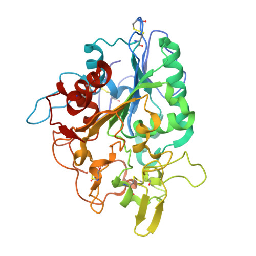
    The crystal structure of poly(3-hydroxybutyrate) (PHB) depolymerase PhaZ7 purified from Paucimonas lemoignei was determined at 1.90 A resolution. The structure consists of a single domain with an alpha/beta hydrolase fold in its core. The active site is analogous to that of serine esterases/lipases and is characterized by the presence of a catalytic triad comprising Ser136, Asp242, and His306. Comparison with other structures in the Protein Data Bank showed a high level of similarity with the Bacillus subtilis lipase LipA (RMSD, 1.55 A). Structural comparison with Penicillium funiculosum PHB depolymerase, the only PHB depolymerase whose structure is already known, revealed significant differences, resulting in an RMSD of 2.80-3.58 A. The two enzymes appear to utilize different types of solvent-exposed residues for biopolymer binding, with aliphatic and hydroxyl residues used in P. funiculosum PHB depolymerase and aromatic residues in PhaZ7. Moreover, the active site of P. funiculosum PHB depolymerase is accessible to the substrate in contrast to the active site of PhaZ7, which is buried. Hence, considerable conformational changes are required in PhaZ7 for the creation of a channel leading to the active site. Taken together, the structural data suggest that PhaZ7 and P. funiculosum PHB depolymerase have adopted different strategies for effective substrate binding in response to their diverse substrate specificity and the lack of a substrate-binding domain.


  • Organizational Affiliation

    Turku Centre for Biotechnology, University of Turku and Abo Akademi University, BioCity, Turku 20521, Finland. tassos.papageorgiou@btk.fi


Macromolecules
Find similar proteins by:  (by identity cutoff)  |  3D Structure
Entity ID: 1
MoleculeChains Sequence LengthOrganismDetailsImage
PHB depolymerase PhaZ7
A, B
342Paucimonas lemoigneiMutation(s): 0 
UniProt
Find proteins for Q939Q9 (Paucimonas lemoignei)
Explore Q939Q9 
Go to UniProtKB:  Q939Q9
Entity Groups  
Sequence Clusters30% Identity50% Identity70% Identity90% Identity95% Identity100% Identity
UniProt GroupQ939Q9
Sequence Annotations
Expand
  • Reference Sequence
Small Molecules
Ligands 1 Unique
IDChains Name / Formula / InChI Key2D Diagram3D Interactions
GOL
Query on GOL

Download Ideal Coordinates CCD File 
C [auth A]GLYCEROL
C3 H8 O3
PEDCQBHIVMGVHV-UHFFFAOYSA-N
Experimental Data & Validation

Experimental Data

  • Method: X-RAY DIFFRACTION
  • Resolution: 1.90 Å
  • R-Value Free: 0.188 
  • R-Value Work: 0.142 
  • R-Value Observed: 0.145 
  • Space Group: C 1 2 1
Unit Cell:
Length ( Å )Angle ( ˚ )
a = 176.383α = 90
b = 46.38β = 99.78
c = 85.779γ = 90
Software Package:
Software NamePurpose
REFMACrefinement
SHARPphasing

Structure Validation

View Full Validation Report


Entry History 

Deposition Data

Revision History  (Full details and data files)

  • Version 1.0: 2008-08-26
    Type: Initial release
  • Version 1.1: 2011-05-08
    Changes: Version format compliance
  • Version 1.2: 2011-07-13
    Changes: Version format compliance
  • Version 1.3: 2018-12-05
    Changes: Data collection, Database references, Refinement description, Source and taxonomy, Structure summary
  • Version 1.4: 2019-01-30
    Changes: Data collection, Experimental preparation
  • Version 1.5: 2019-07-10
    Changes: Data collection
  • Version 1.6: 2019-07-24
    Changes: Data collection