2VBK

NATIVE TAILSPIKE PROTEIN OF BACTERIOPHAGE SF6


Experimental Data Snapshot

  • Method: X-RAY DIFFRACTION
  • Resolution: 1.25 Å
  • R-Value Free: 0.142 
  • R-Value Work: 0.119 
  • R-Value Observed: 0.120 

wwPDB Validation   3D Report Full Report


This is version 2.5 of the entry. See complete history


Literature

An intersubunit active site between supercoiled parallel beta helices in the trimeric tailspike endorhamnosidase of Shigella flexneri Phage Sf6.

Muller, J.J.Barbirz, S.Heinle, K.Freiberg, A.Seckler, R.Heinemann, U.

(2008) Structure 16: 766-775

  • DOI: https://doi.org/10.1016/j.str.2008.01.019
  • Primary Citation of Related Structures:  
    2VBE, 2VBK, 2VBM

  • PubMed Abstract: 

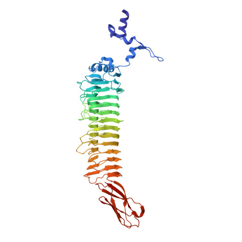
    Sf6 belongs to the Podoviridae family of temperate bacteriophages that infect gram-negative bacteria by insertion of their double-stranded DNA. They attach to their hosts specifically via their tailspike proteins. The 1.25 A crystal structure of Shigella phage Sf6 tailspike protein (Sf6 TSP) reveals a conserved architecture with a central, right-handed beta helix. In the trimer of Sf6 TSP, the parallel beta helices form a left-handed, coiled-beta coil with a pitch of 340 A. The C-terminal domain consists of a beta sandwich reminiscent of viral capsid proteins. Further crystallographic and biochemical analyses show a Shigella cell wall O-antigen fragment to bind to an endorhamnosidase active site located between two beta-helix subunits each anchoring one catalytic carboxylate. The functionally and structurally related bacteriophage, P22 TSP, lacks sequence identity with Sf6 TSP and has its active sites on single subunits. Sf6 TSP may serve as an example for the evolution of different host specificities on a similar general architecture.


  • Organizational Affiliation

    Max-Delbrück-Centrum für Molekulare Medizin, Robert-Rössle-Str. 10, 13125 Berlin, Germany.


Macromolecules
Find similar proteins by:  (by identity cutoff)  |  3D Structure
Entity ID: 1
MoleculeChains Sequence LengthOrganismDetailsImage
TAILSPIKE-PROTEIN514Lederbergvirus Sf6Mutation(s): 0 
UniProt
Find proteins for Q9XJP3 (Shigella phage Sf6)
Explore Q9XJP3 
Go to UniProtKB:  Q9XJP3
Entity Groups  
Sequence Clusters30% Identity50% Identity70% Identity90% Identity95% Identity100% Identity
UniProt GroupQ9XJP3
Sequence Annotations
Expand
  • Reference Sequence
Small Molecules
Ligands 6 Unique
IDChains Name / Formula / InChI Key2D Diagram3D Interactions
PO4
Query on PO4

Download Ideal Coordinates CCD File 
U [auth A],
V [auth A],
W [auth A]
PHOSPHATE ION
O4 P
NBIIXXVUZAFLBC-UHFFFAOYSA-K
GOL
Query on GOL

Download Ideal Coordinates CCD File 
O [auth A]
P [auth A]
Q [auth A]
R [auth A]
S [auth A]
O [auth A],
P [auth A],
Q [auth A],
R [auth A],
S [auth A],
T [auth A]
GLYCEROL
C3 H8 O3
PEDCQBHIVMGVHV-UHFFFAOYSA-N
PGR
Query on PGR

Download Ideal Coordinates CCD File 
M [auth A],
N [auth A]
R-1,2-PROPANEDIOL
C3 H8 O2
DNIAPMSPPWPWGF-GSVOUGTGSA-N
EDO
Query on EDO

Download Ideal Coordinates CCD File 
AA [auth A]
BA [auth A]
D [auth A]
E [auth A]
F [auth A]
AA [auth A],
BA [auth A],
D [auth A],
E [auth A],
F [auth A],
G [auth A],
H [auth A],
I [auth A],
J [auth A],
K [auth A],
L [auth A],
Z [auth A]
1,2-ETHANEDIOL
C2 H6 O2
LYCAIKOWRPUZTN-UHFFFAOYSA-N
CO2
Query on CO2

Download Ideal Coordinates CCD File 
X [auth A],
Y [auth A]
CARBON DIOXIDE
C O2
CURLTUGMZLYLDI-UHFFFAOYSA-N
MG
Query on MG

Download Ideal Coordinates CCD File 
B [auth A],
C [auth A]
MAGNESIUM ION
Mg
JLVVSXFLKOJNIY-UHFFFAOYSA-N
Experimental Data & Validation

Experimental Data

  • Method: X-RAY DIFFRACTION
  • Resolution: 1.25 Å
  • R-Value Free: 0.142 
  • R-Value Work: 0.119 
  • R-Value Observed: 0.120 
  • Space Group: H 3
Unit Cell:
Length ( Å )Angle ( ˚ )
a = 96.231α = 90
b = 96.231β = 90
c = 182.433γ = 120
Software Package:
Software NamePurpose
REFMACrefinement
DENZOdata reduction
SCALEPACKdata scaling
AMoREphasing

Structure Validation

View Full Validation Report


Entry History 

Deposition Data

Revision History  (Full details and data files)

  • Version 1.0: 2008-04-01
    Type: Initial release
  • Version 1.1: 2011-05-08
    Changes: Version format compliance
  • Version 1.2: 2011-07-13
    Changes: Version format compliance
  • Version 2.0: 2018-12-19
    Changes: Atomic model, Data collection, Database references, Derived calculations
  • Version 2.1: 2019-01-30
    Changes: Data collection, Experimental preparation
  • Version 2.2: 2019-05-22
    Changes: Data collection, Experimental preparation
  • Version 2.3: 2019-07-10
    Changes: Data collection
  • Version 2.4: 2019-07-24
    Changes: Data collection
  • Version 2.5: 2023-12-13
    Changes: Data collection, Database references, Derived calculations, Other, Refinement description