2VAT

Crystal structure of deacetylcephalosporin C acetyltransferase in complex with coenzyme A


Experimental Data Snapshot

  • Method: X-RAY DIFFRACTION
  • Resolution: 2.20 Å
  • R-Value Free: 0.230 
  • R-Value Work: 0.200 
  • R-Value Observed: 0.201 

wwPDB Validation   3D Report Full Report


This is version 1.3 of the entry. See complete history


Literature

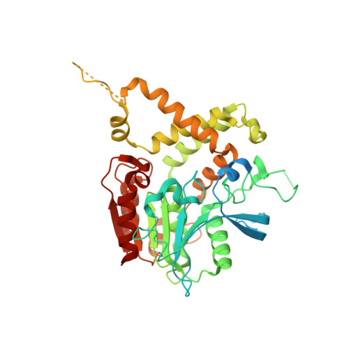
The Last Step in Cephalosporin C Formation Revealed: Crystal Structures of Deacetylcephalosporin C Acetyltransferase from Acremonium Chrysogenum in Complexes with Reaction Intermediates.

Lejon, S.Ellis, J.Valegard, K.

(2008) J Mol Biol 377: 935

  • DOI: https://doi.org/10.1016/j.jmb.2008.01.047
  • Primary Citation of Related Structures:  
    2VAT, 2VAV, 2VAX

  • PubMed Abstract: 

    Deacetylcephalosporin C acetyltransferase (DAC-AT) catalyses the last step in the biosynthesis of cephalosporin C, a broad-spectrum beta-lactam antibiotic of large clinical importance. The acetyl transfer step has been suggested to be limiting for cephalosporin C biosynthesis, but has so far escaped detailed structural analysis. We present here the crystal structures of DAC-AT in complexes with reaction intermediates, providing crystallographic snapshots of the reaction mechanism. The enzyme is found to belong to the alpha/beta hydrolase class of acetyltransferases, and the structures support previous observations of a double displacement mechanism for the acetyl transfer reaction in other members of this class of enzymes. The structures of DAC-AT reported here provide evidence of a stable acyl-enzyme complex, thus underpinning a mechanism involving acetylation of a catalytic serine residue by acetyl coenzyme A, followed by transfer of the acetyl group to deacetylcephalosporin C through a suggested tetrahedral transition state.


  • Organizational Affiliation

    Department of Cell and Molecular Biology, Uppsala University, Biomedical Centre, Box 596, S-751 24 Uppsala, Sweden.


Macromolecules
Find similar proteins by:  (by identity cutoff)  |  3D Structure
Entity ID: 1
MoleculeChains Sequence LengthOrganismDetailsImage
ACETYL-COA--DEACETYLCEPHALOSPORIN C ACETYLTRANSFERASE
A, B, C, D, E
A, B, C, D, E, F, G, H, I, J, K, L
444Hapsidospora chrysogenaMutation(s): 0 
EC: 2.3.1.175
UniProt
Find proteins for P39058 (Hapsidospora chrysogena)
Explore P39058 
Go to UniProtKB:  P39058
Entity Groups  
Sequence Clusters30% Identity50% Identity70% Identity90% Identity95% Identity100% Identity
UniProt GroupP39058
Sequence Annotations
Expand
  • Reference Sequence
Small Molecules
Ligands 3 Unique
IDChains Name / Formula / InChI Key2D Diagram3D Interactions
COA
Query on COA

Download Ideal Coordinates CCD File 
BA [auth H]
DA [auth I]
GA [auth J]
IA [auth K]
KA [auth L]
BA [auth H],
DA [auth I],
GA [auth J],
IA [auth K],
KA [auth L],
M [auth A],
O [auth B],
R [auth C],
T [auth D],
V [auth E],
X [auth F],
Z [auth G]
COENZYME A
C21 H36 N7 O16 P3 S
RGJOEKWQDUBAIZ-IBOSZNHHSA-N
GOL
Query on GOL

Download Ideal Coordinates CCD File 
EA [auth I],
P [auth B]
GLYCEROL
C3 H8 O3
PEDCQBHIVMGVHV-UHFFFAOYSA-N
ACT
Query on ACT

Download Ideal Coordinates CCD File 
AA [auth G]
CA [auth H]
FA [auth I]
HA [auth J]
JA [auth K]
AA [auth G],
CA [auth H],
FA [auth I],
HA [auth J],
JA [auth K],
LA [auth L],
N [auth A],
Q [auth B],
S [auth C],
U [auth D],
W [auth E],
Y [auth F]
ACETATE ION
C2 H3 O2
QTBSBXVTEAMEQO-UHFFFAOYSA-M
Experimental Data & Validation

Experimental Data

  • Method: X-RAY DIFFRACTION
  • Resolution: 2.20 Å
  • R-Value Free: 0.230 
  • R-Value Work: 0.200 
  • R-Value Observed: 0.201 
  • Space Group: P 1 21 1
Unit Cell:
Length ( Å )Angle ( ˚ )
a = 121.959α = 90
b = 109.278β = 90.23
c = 197.001γ = 90
Software Package:
Software NamePurpose
MOSFLMdata reduction
SCALAdata scaling
SHELXphasing
autoSHARPphasing
REFMACrefinement

Structure Validation

View Full Validation Report


Entry History 

Deposition Data

Revision History  (Full details and data files)

  • Version 1.0: 2008-09-23
    Type: Initial release
  • Version 1.1: 2011-05-08
    Changes: Version format compliance
  • Version 1.2: 2011-07-13
    Changes: Version format compliance
  • Version 1.3: 2018-01-17
    Changes: Data collection