2R2O

Crystal structure of the effector domain of human Plexin B1


Experimental Data Snapshot

  • Method: X-RAY DIFFRACTION
  • Resolution: 2.00 Å
  • R-Value Free: 0.269 
  • R-Value Work: 0.222 
  • R-Value Observed: 0.224 

wwPDB Validation   3D Report Full Report


This is version 1.2 of the entry. See complete history


Literature

Binding of Rac1, Rnd1, and RhoD to a novel Rho GTPase interaction motif destabilizes dimerization of the plexin-B1 effector domain.

Tong, Y.Chugha, P.Hota, P.K.Alviani, R.S.Li, M.Tempel, W.Shen, L.Park, H.W.Buck, M.

(2007) J Biol Chem 282: 37215-37224

  • DOI: https://doi.org/10.1074/jbc.M703800200
  • Primary Citation of Related Structures:  
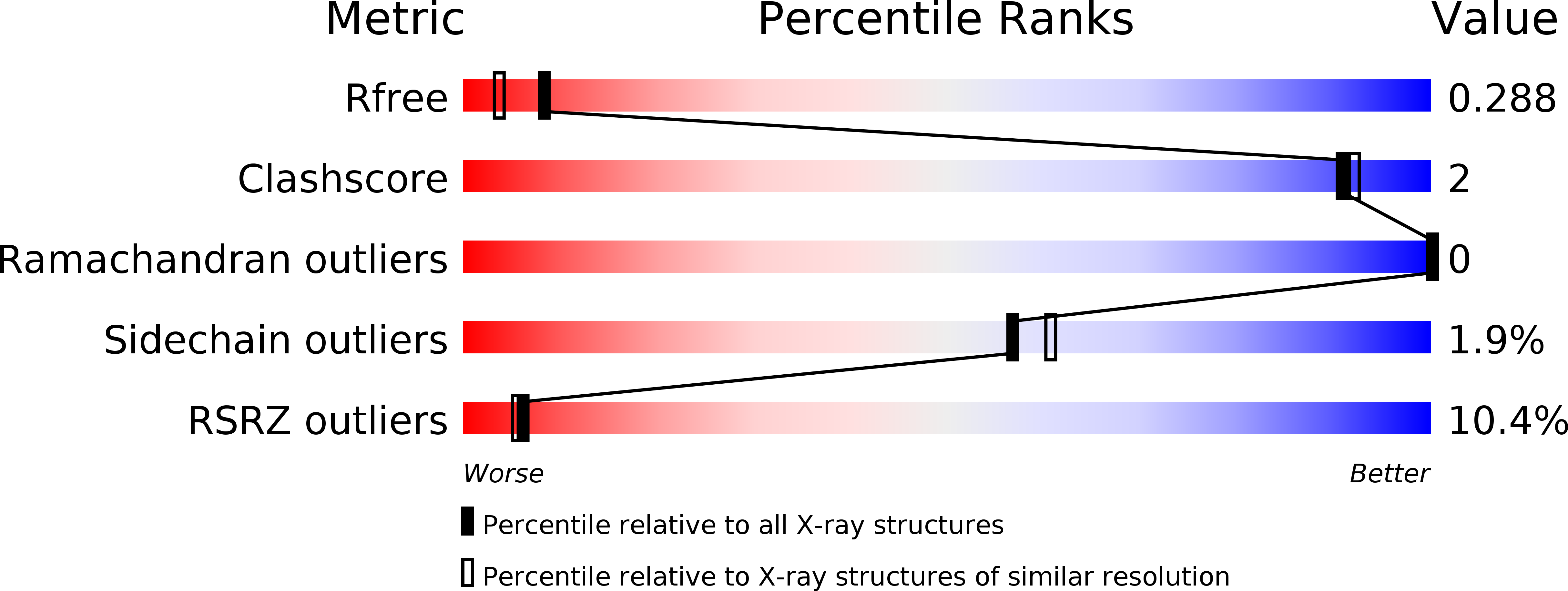
    2R2O

  • PubMed Abstract: 

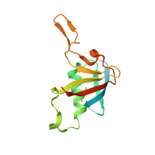
    Plexins are the first known transmembrane receptors that interact directly with small GTPases. On binding to certain Rho family GTPases, the receptor regulates the remodeling of the actin cytoskeleton and alters cell movement in response to semaphorin guidance cues. In a joint solution NMR spectroscopy and x-ray crystallographic study, we characterize a 120-residue cytoplasmic independent folding domain of plexin-B1 that directly binds three Rho family GTPases, Rac1, Rnd1, and RhoD. The NMR data show that, surprisingly, the Cdc42/Rac interactive binding-like motif of plexin-B1 is not involved in this interaction. Instead, all three GTPases interact with the same region, beta-strands 3 and 4 and a short alpha-helical segment of the plexin domain. The 2.0 A resolution x-ray structure shows that these segments are brought together by the tertiary structure of the ubiquitin-like fold. In the crystal, the protein is dimerized with C2 symmetry through a four-stranded antiparallel beta-sheet that is formed outside the fold by a long loop between the monomers. This region is adjacent to the GTPase binding motifs identified by NMR. Destabilization of the dimer in solution by binding of any one of the three GTPases suggests a model for receptor regulation that involves bidirectional signaling. The model implies a multifunctional role for the GTPase-plexin interaction that includes conformational change and a localization of active receptors in the signaling mechanism.


  • Organizational Affiliation

    Department of Physiology, Case Western Reserve University School of Medicine, Cleveland, Ohio 44106, USA.


Macromolecules
Find similar proteins by:  (by identity cutoff)  |  3D Structure
Entity ID: 1
MoleculeChains Sequence LengthOrganismDetailsImage
Plexin-B1
A, B
138Homo sapiensMutation(s): 0 
Gene Names: PLXNB1KIAA0407PLXN5SEP
UniProt & NIH Common Fund Data Resources
Find proteins for O43157 (Homo sapiens)
Explore O43157 
Go to UniProtKB:  O43157
PHAROS:  O43157
GTEx:  ENSG00000164050 
Entity Groups  
Sequence Clusters30% Identity50% Identity70% Identity90% Identity95% Identity100% Identity
UniProt GroupO43157
Sequence Annotations
Expand
  • Reference Sequence
Small Molecules
Ligands 1 Unique
IDChains Name / Formula / InChI Key2D Diagram3D Interactions
UNX
Query on UNX

Download Ideal Coordinates CCD File 
C [auth A]
D [auth A]
E [auth A]
F [auth A]
G [auth A]
C [auth A],
D [auth A],
E [auth A],
F [auth A],
G [auth A],
H [auth A],
I [auth A],
J [auth B],
K [auth B],
L [auth B],
M [auth B],
N [auth B]
UNKNOWN ATOM OR ION
X
Modified Residues  1 Unique
IDChains TypeFormula2D DiagramParent
MSE
Query on MSE
A, B
L-PEPTIDE LINKINGC5 H11 N O2 SeMET
Experimental Data & Validation

Experimental Data

  • Method: X-RAY DIFFRACTION
  • Resolution: 2.00 Å
  • R-Value Free: 0.269 
  • R-Value Work: 0.222 
  • R-Value Observed: 0.224 
  • Space Group: P 21 21 21
Unit Cell:
Length ( Å )Angle ( ˚ )
a = 50.979α = 90
b = 56.571β = 90
c = 99.366γ = 90
Software Package:
Software NamePurpose
DENZOdata reduction
SCALEPACKdata scaling
SHELXphasing
RESOLVEphasing
REFMACrefinement
PDB_EXTRACTdata extraction
ADSCdata collection

Structure Validation

View Full Validation Report


Entry History 

Revision History  (Full details and data files)

  • Version 1.0: 2007-09-04
    Type: Initial release
  • Version 1.1: 2011-07-13
    Changes: Advisory, Version format compliance
  • Version 1.2: 2017-10-25
    Changes: Refinement description