2QB1

2TEL crystallization module


Experimental Data Snapshot

  • Method: X-RAY DIFFRACTION
  • Resolution: 2.61 Å
  • R-Value Free: 0.241 
  • R-Value Work: 0.211 
  • R-Value Observed: 0.212 

wwPDB Validation   3D Report Full Report


This is version 1.4 of the entry. See complete history


Literature

Polymer-driven crystallization.

Nauli, S.Farr, S.Lee, Y.J.Kim, H.Y.Faham, S.Bowie, J.U.

(2007) Protein Sci 16: 2542-2551

  • DOI: https://doi.org/10.1110/ps.073074207
  • Primary Citation of Related Structures:  
    2QAR, 2QB0, 2QB1

  • PubMed Abstract: 

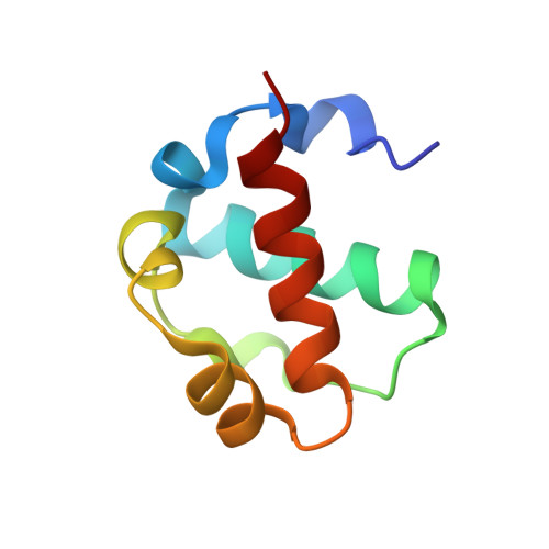
    Obtaining well-diffracting crystals of macromolecules remains a significant barrier to structure determination. Here we propose and test a new approach to crystallization, in which the crystallization target is fused to a polymerizing protein module, so that polymer formation drives crystallization of the target. We test the approach using a polymerization module called 2TEL, which consists of two tandem sterile alpha motif (SAM) domains from the protein translocation Ets leukemia (TEL). The 2TEL module is engineered to polymerize as the pH is lowered, which allows the subtle modulation of polymerization needed for crystal formation. We show that the 2TEL module can drive the crystallization of 11 soluble proteins, including three that resisted prior crystallization attempts. In addition, the 2TEL module crystallizes in the presence of various detergents, suggesting that it might facilitate membrane protein crystallization. The crystal structures of two fusion proteins show that the TELSAM polymer is responsible for the majority of contacts in the crystal lattice. The results suggest that biological polymers could be designed as crystallization modules.


  • Organizational Affiliation

    UCLA-DOE Institute of Genomics and Proteomics, University of California, Los Angeles 90095-1570, USA.


Macromolecules
Find similar proteins by:  (by identity cutoff)  |  3D Structure
Entity ID: 1
MoleculeChains Sequence LengthOrganismDetailsImage
E80-TELSAM domain
A, B
78Escherichia coliMutation(s): 0 
UniProt & NIH Common Fund Data Resources
Find proteins for P41212 (Homo sapiens)
Explore P41212 
Go to UniProtKB:  P41212
PHAROS:  P41212
GTEx:  ENSG00000139083 
Entity Groups  
Sequence Clusters30% Identity50% Identity70% Identity90% Identity95% Identity100% Identity
UniProt GroupP41212
Sequence Annotations
Expand
  • Reference Sequence
Experimental Data & Validation

Experimental Data

  • Method: X-RAY DIFFRACTION
  • Resolution: 2.61 Å
  • R-Value Free: 0.241 
  • R-Value Work: 0.211 
  • R-Value Observed: 0.212 
  • Space Group: P 31
Unit Cell:
Length ( Å )Angle ( ˚ )
a = 65.686α = 90
b = 65.686β = 90
c = 36.144γ = 120
Software Package:
Software NamePurpose
DENZOdata reduction
SCALEPACKdata scaling
PHASERphasing
REFMACrefinement
PDB_EXTRACTdata extraction

Structure Validation

View Full Validation Report


Entry History 

Deposition Data

Revision History  (Full details and data files)

  • Version 1.0: 2008-10-14
    Type: Initial release
  • Version 1.1: 2011-07-13
    Changes: Advisory, Version format compliance
  • Version 1.2: 2017-10-18
    Changes: Refinement description
  • Version 1.3: 2020-06-24
    Changes: Database references, Source and taxonomy
  • Version 1.4: 2023-08-30
    Changes: Data collection, Database references, Refinement description