Analysis of coiled-coil interactions between core proteins of the spindle pole body.
Zizlsperger, N., Malashkevich, V.N., Pillay, S., Keating, A.E.(2008) Biochemistry 47: 11858-11868
- PubMed: 18850724
- DOI: https://doi.org/10.1021/bi801378z
- Primary Citation of Related Structures:
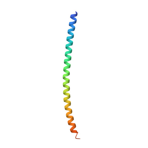
2Q6Q - PubMed Abstract:
The spindle pole body (SPB) is a multiprotein complex that organizes microtubules in yeast. Due to its large size and association with the nuclear membrane, little is known about its detailed structure. In particular, although many SPB components and some of the interactions between them have been identified, the molecular details of how most of these interactions occur are not known. The prevalence of predicted coiled-coil regions in SPB proteins suggests that some interactions may occur via coiled coils. Here this hypothesis is supported by biochemical characterization of isolated coiled-coil peptides derived from SPB proteins. Formation of four strongly self-associating coiled-coil complexes from Spc29, Spc42, and Spc72 was demonstrated by circular dichroism (CD) spectroscopy and a fluorescence resonance energy transfer (FRET) assay. Many weaker self- and heteroassociations were also detected by CD, FRET, and/or cross-linking. The thermal stabilities of nine candidate homooligomers were assessed; six unfolded cooperatively with melting temperatures ranging from <11 to >50 degrees C. Solution studies established that coiled-coil peptides derived from Spc42 and Spc72 form parallel dimers, and this was confirmed for Spc42 by a high-resolution crystal structure. These data contribute to a growing body of knowledge that will ultimately provide a detailed model of the SPB structure.
Organizational Affiliation:
Department of Biology, Massachusetts Institute of Technology, 77 Massachusetts Avenue, Cambridge, Massachusetts 02139, USA.