2Q33

Crystal structure of all-D monellin at 1.8 A resolution


Experimental Data Snapshot

  • Method: X-RAY DIFFRACTION
  • Resolution: 1.80 Å
  • R-Value Free: 0.215 
  • R-Value Work: 0.175 
  • R-Value Observed: 0.175 

wwPDB Validation   3D Report Full Report


This is version 1.2 of the entry. See complete history


Literature

Structure of an Enantiomeric Protein, D-Monellin at 1.8 A Resolution.

Hung, L.-W.Kohmura, M.Ariyoshi, Y.Kim, S.-H.

(1998) Acta Crystallogr D Biol Crystallogr 54: 494-500

  • DOI: https://doi.org/10.1107/s0907444997012225
  • Primary Citation of Related Structures:  
    2Q33

  • PubMed Abstract: 

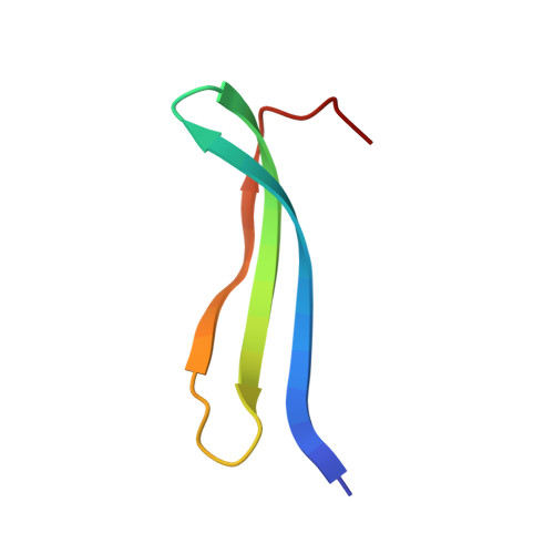
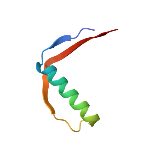
    The D-enantiomer of a potently sweet protein, monellin, has been crystallized and analyzed by X-ray crystallography at 1.8 A resolut ion. Two crystal forms (I and II) appeared under crystallization conditions similar, but not identical, to the crystallization conditions of natural L-monellin. There are four molecules per asymmetric unit in crystal form I and one in crystal form II. Crystal form I is not reproducible and is equivalent to that of monoclinic L-monellin. Intermonomer contacts in crystal form II are very different from those found in natural L-monellin crystals. The backbone trace of D-monellin resembles very closely the mirror image of that of L-monellin, but the N- and C-terminus backbones as well as several side-chain conformations of D-monellin are different from those of natural L-monellin. Most of these apparent differences may be attributable to the crystal packing differences.


  • Organizational Affiliation

    Graduate Group in Biophysics, Department of Chemistry, Univesity of California, Berkeley, 94720, USA.


Macromolecules
Find similar proteins by:  (by identity cutoff)  |  3D Structure
Entity ID: 1
MoleculeChains Sequence LengthOrganismDetailsImage
D-MONELLIN CHAIN A43N/AMutation(s): 0 
Entity Groups  
Sequence Clusters30% Identity50% Identity70% Identity90% Identity95% Identity100% Identity
Sequence Annotations
Expand
  • Reference Sequence
Find similar proteins by:  (by identity cutoff)  |  3D Structure
Entity ID: 2
MoleculeChains Sequence LengthOrganismDetailsImage
D-MONELLIN CHAIN B48N/AMutation(s): 0 
Entity Groups  
Sequence Clusters30% Identity50% Identity70% Identity90% Identity95% Identity100% Identity
Sequence Annotations
Expand
  • Reference Sequence
Experimental Data & Validation

Experimental Data

  • Method: X-RAY DIFFRACTION
  • Resolution: 1.80 Å
  • R-Value Free: 0.215 
  • R-Value Work: 0.175 
  • R-Value Observed: 0.175 
  • Space Group: P 1 2 1
Unit Cell:
Length ( Å )Angle ( ˚ )
a = 40.4α = 90
b = 32.94β = 96.73
c = 41.11γ = 90
Software Package:
Software NamePurpose
AMoREphasing
X-PLORrefinement
MAR345data collection
SCALEPACKdata scaling

Structure Validation

View Full Validation Report


Entry History 

Deposition Data

Revision History  (Full details and data files)

  • Version 1.0: 2007-11-13
    Type: Initial release
  • Version 1.1: 2011-07-13
    Changes: Version format compliance
  • Version 1.2: 2017-10-18
    Changes: Refinement description