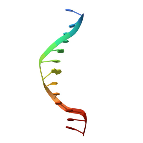
2OXV

Structure of the A138T promiscuous mutant of the EcoRI restriction endonuclease bound to its cognate recognition site.


Experimental Data Snapshot

  • Method: X-RAY DIFFRACTION
  • Resolution: 1.95 Å
  • R-Value Free: 0.244 
  • R-Value Work: 0.202 

wwPDB Validation   3D Report Full Report


This is version 1.3 of the entry. See complete history


Literature

Structural and thermodynamic basis for enhanced DNA binding by a promiscuous mutant EcoRI endonuclease.

Sapienza, P.J.Rosenberg, J.M.Jen-Jacobson, L.

(2007) Structure 15: 1368-1382

  • DOI: https://doi.org/10.1016/j.str.2007.09.014
  • Primary Citation of Related Structures:  
    2OXV

  • PubMed Abstract: 

    Promiscuous mutant EcoRI endonucleases bind to the canonical site GAATTC more tightly than does the wild-type endonuclease, yet cleave variant (EcoRI(*)) sites more rapidly than does wild-type. The crystal structure of the A138T promiscuous mutant homodimer in complex with a GAATTC site is nearly identical to that of the wild-type complex, except that the Thr138 side chains make packing interactions with bases in the 5'-flanking regions outside the recognition hexanucleotide while excluding two bound water molecules seen in the wild-type complex. Molecular dynamics simulations confirm exclusion of these waters. The structure and simulations suggest possible reasons why binding of the A138T protein to the GAATTC site has DeltaS degrees more favorable and DeltaH degrees less favorable than for wild-type endonuclease binding. The interactions of Thr138 with flanking bases may permit A138T, unlike wild-type enzyme, to form complexes with EcoRI(*) sites that structurally resemble the specific wild-type complex with GAATTC.


  • Organizational Affiliation

    Department of Biological Sciences, University of Pittsburgh, Pittsburgh, PA 15260, USA.


Macromolecules

Find similar proteins by:  (by identity cutoff)  |  3D Structure
Entity ID: 2
MoleculeChains Sequence LengthOrganismDetailsImage
Type II restriction enzyme EcoRIB [auth A]277Escherichia coliMutation(s): 0 
Gene Names: ecoRIR
EC: 3.1.21.4
UniProt
Find proteins for P00642 (Escherichia coli)
Explore P00642 
Go to UniProtKB:  P00642
Entity Groups  
Sequence Clusters30% Identity50% Identity70% Identity90% Identity95% Identity100% Identity
UniProt GroupP00642
Sequence Annotations
Expand
  • Reference Sequence

Find similar nucleic acids by:  Sequence   |   3D Structure  

Entity ID: 1
MoleculeChains LengthOrganismImage
DNA (5'-D(*TP*CP*GP*CP*GP*AP*AP*TP*TP*CP*GP*CP*G)-3')A [auth C]13N/A
Sequence Annotations
Expand
  • Reference Sequence
Experimental Data & Validation

Experimental Data

  • Method: X-RAY DIFFRACTION
  • Resolution: 1.95 Å
  • R-Value Free: 0.244 
  • R-Value Work: 0.202 
  • Space Group: P 3 2 1
Unit Cell:
Length ( Å )Angle ( ˚ )
a = 116.473α = 90
b = 116.473β = 90
c = 48.476γ = 120
Software Package:
Software NamePurpose
DENZOdata reduction
SCALEPACKdata scaling
CNSrefinement
PDB_EXTRACTdata extraction
ADSCdata collection
HKL-2000data reduction
CNSphasing

Structure Validation

View Full Validation Report


Entry History 

Deposition Data

Revision History  (Full details and data files)

  • Version 1.0: 2007-10-23
    Type: Initial release
  • Version 1.1: 2011-07-13
    Changes: Version format compliance
  • Version 1.2: 2017-10-18
    Changes: Refinement description
  • Version 1.3: 2023-08-30
    Changes: Data collection, Database references, Refinement description