2OHY

X-ray Crystal Structure of Tyrosine Aminomutase from streptomyces globisporus


Experimental Data Snapshot

  • Method: X-RAY DIFFRACTION
  • Resolution: 2.50 Å
  • R-Value Free: 0.263 
  • R-Value Work: 0.192 
  • R-Value Observed: 0.199 

wwPDB Validation   3D Report Full Report


This is version 2.0 of the entry. See complete history


Literature

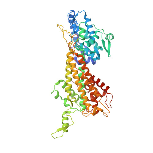
The structure of L-tyrosine 2,3-aminomutase from the C-1027 enediyne antitumor antibiotic biosynthetic pathway

Christianson, C.V.Montavon, T.J.Van Lanen, S.G.Shen, B.Bruner, S.D.

(2007) Biochemistry 46: 7205-7214

  • DOI: https://doi.org/10.1021/bi7003685
  • Primary Citation of Related Structures:  
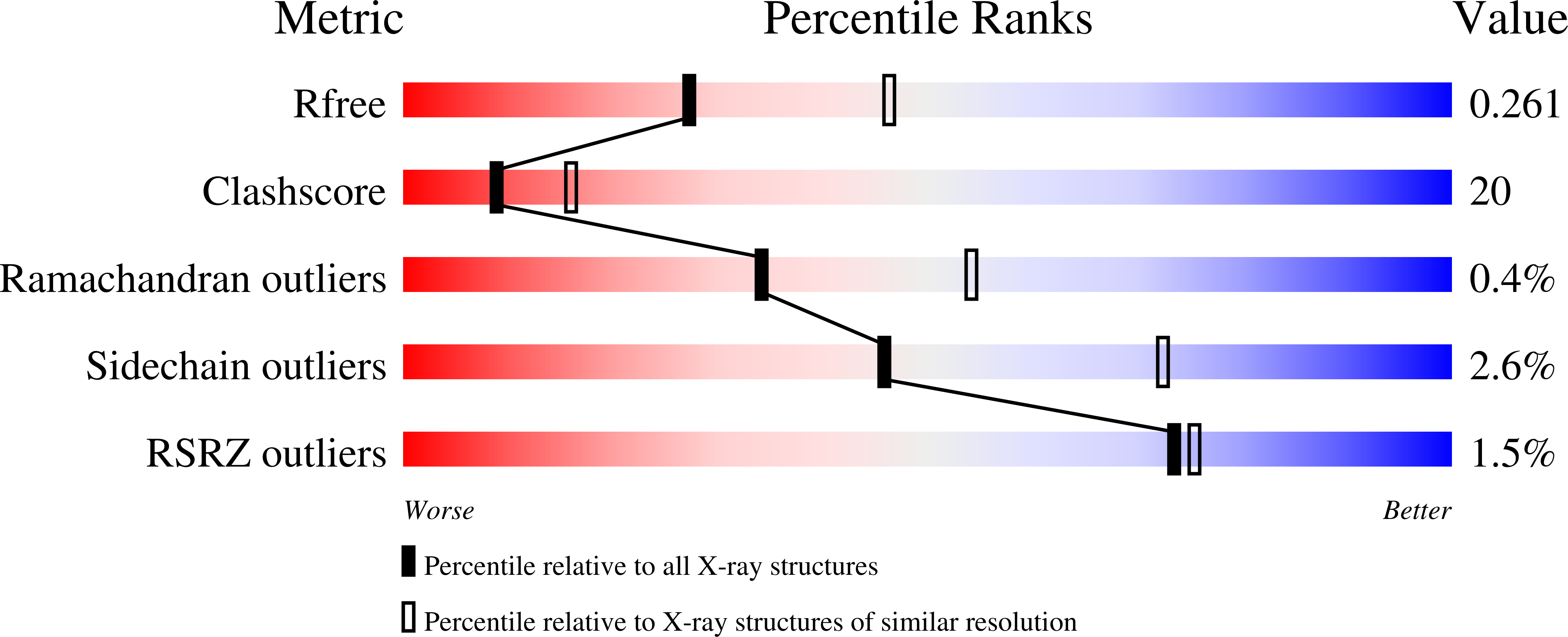
    2OHY

  • PubMed Abstract: 

    The SgcC4 l-tyrosine 2,3-aminomutase (SgTAM) catalyzes the formation of (S)-beta-tyrosine in the biosynthetic pathway of the enediyne antitumor antibiotic C-1027. SgTAM is homologous to the histidine ammonia lyase family of enzymes whose activity is dependent on the methylideneimidazole-5-one (MIO) cofactor. Unlike the lyase enzymes, SgTAM catalyzes additional chemical transformations resulting in an overall stereospecific 1,2-amino shift in the substrate l-tyrosine to generate (S)-beta-tyrosine. Previously, we provided kinetic, spectroscopic, and mutagenesis data supporting the presence of MIO in the active site of SgTAM [Christenson, S. D.; Wu, W.; Spies, A.; Shen, B.; and Toney, M. D. (2003) Biochemistry 42, 12708-12718]. Here we report the first X-ray crystal structure of an MIO-containing aminomutase, SgTAM, and confirm the structural homology of SgTAM to ammonia lyases. Comparison of the structure of SgTAM to the l-tyrosine ammonia lyase from Rhodobacter sphaeroides provides insight into the structural basis for aminomutase activity. The results show that SgTAM has a closed active site well suited to retain ammonia and minimize the formation of lyase elimination products. The amino acid determinants for substrate recognition and catalysis can be predicted from the structure, setting the framework for detailed mechanistic investigations.


  • Organizational Affiliation

    Department of Chemistry, Boston College, Eugene F. Merkert Chemistry Center, Chestnut Hill, Massachusetts 02467, USA.


Macromolecules
Find similar proteins by:  (by identity cutoff)  |  3D Structure
Entity ID: 1
MoleculeChains Sequence LengthOrganismDetailsImage
Tyrosine aminomutase
A, B
539Streptomyces globisporusMutation(s): 1 
EC: 5.4.3.6
UniProt
Find proteins for Q8GMG0 (Streptomyces globisporus)
Explore Q8GMG0 
Go to UniProtKB:  Q8GMG0
Entity Groups  
Sequence Clusters30% Identity50% Identity70% Identity90% Identity95% Identity100% Identity
UniProt GroupQ8GMG0
Sequence Annotations
Expand
  • Reference Sequence
Small Molecules
Modified Residues  1 Unique
IDChains TypeFormula2D DiagramParent
MDO
Query on MDO
A, B
L-PEPTIDE LINKINGC8 H11 N3 O3ALA, SER, GLY
Experimental Data & Validation

Experimental Data

  • Method: X-RAY DIFFRACTION
  • Resolution: 2.50 Å
  • R-Value Free: 0.263 
  • R-Value Work: 0.192 
  • R-Value Observed: 0.199 
  • Space Group: P 21 21 2
Unit Cell:
Length ( Å )Angle ( ˚ )
a = 92.597α = 90
b = 146.178β = 90
c = 75.437γ = 90
Software Package:
Software NamePurpose
DENZOdata reduction
SCALEPACKdata scaling
AMoREphasing
CNSrefinement
PDB_EXTRACTdata extraction
CBASSdata collection
HKL-2000data reduction
HKL-2000data scaling

Structure Validation

View Full Validation Report


Entry History 

Deposition Data

Revision History  (Full details and data files)

  • Version 1.0: 2007-06-12
    Type: Initial release
  • Version 1.1: 2008-05-01
    Changes: Version format compliance
  • Version 1.2: 2011-07-13
    Changes: Derived calculations, Version format compliance
  • Version 1.3: 2017-10-18
    Changes: Refinement description
  • Version 1.4: 2023-08-30
    Changes: Data collection, Database references, Derived calculations, Refinement description
  • Version 2.0: 2023-11-15
    Changes: Atomic model, Data collection, Derived calculations