2OCP

Crystal Structure of Human Deoxyguanosine Kinase


Experimental Data Snapshot

  • Method: X-RAY DIFFRACTION
  • Resolution: 2.80 Å
  • R-Value Free: 0.277 
  • R-Value Work: 0.265 
  • R-Value Observed: 0.265 

wwPDB Validation   3D Report Full Report


Ligand Structure Quality Assessment 


This is version 1.3 of the entry. See complete history


Literature

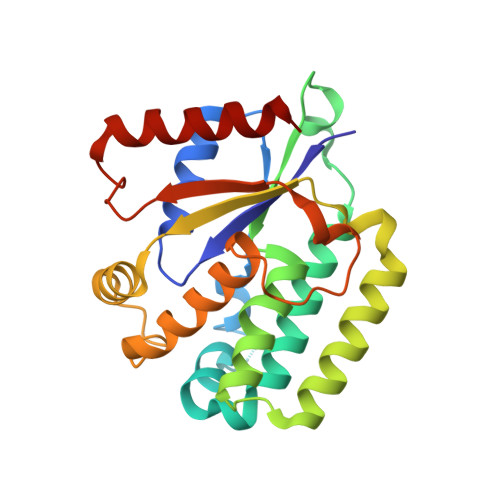
Structural Basis for Substrate Specificities of Cellular Deoxyribonucleoside Kinases.

Johansson, K.Ramaswamy, S.Ljungkrantz, C.Knecht, W.Piskur, J.Munch-Petersen, B.Eriksson, S.Eklund, H.

(2001) Nat Struct Biol 8: 616

  • DOI: https://doi.org/10.1038/89661
  • Primary Citation of Related Structures:  
    1J90, 2OCP

  • PubMed Abstract: 

    Deoxyribonucleoside kinases phosphorylate deoxyribonucleosides and activate a number of medically important nucleoside analogs. Here we report the structure of the Drosophila deoxyribonucleoside kinase with deoxycytidine bound at the nucleoside binding site and that of the human deoxyguanosine kinase with ATP at the nucleoside substrate binding site. Compared to the human kinase, the Drosophila kinase has a wider substrate cleft, which may be responsible for the broad substrate specificity of this enzyme. The human deoxyguanosine kinase is highly specific for purine substrates; this is apparently due to the presence of Arg 118, which provides favorable hydrogen bonding interactions with the substrate. The two new structures provide an explanation for the substrate specificity of cellular deoxyribonucleoside kinases.


  • Organizational Affiliation

    Department of Molecular Biology, Swedish University of Agricultural Sciences, Box 590, Biomedical Center, S-751 24 Uppsala, Sweden.


Macromolecules
Find similar proteins by:  (by identity cutoff)  |  3D Structure
Entity ID: 1
MoleculeChains Sequence LengthOrganismDetailsImage
Deoxyguanosine kinase
A, B, C, D, E
A, B, C, D, E, F, G, H
241Homo sapiensMutation(s): 1 
Gene Names: DGUOKDGK
EC: 2.7.1.113
UniProt & NIH Common Fund Data Resources
Find proteins for Q16854 (Homo sapiens)
Explore Q16854 
Go to UniProtKB:  Q16854
PHAROS:  Q16854
GTEx:  ENSG00000114956 
Entity Groups  
Sequence Clusters30% Identity50% Identity70% Identity90% Identity95% Identity100% Identity
UniProt GroupQ16854
Sequence Annotations
Expand
  • Reference Sequence
Experimental Data & Validation

Experimental Data

  • Method: X-RAY DIFFRACTION
  • Resolution: 2.80 Å
  • R-Value Free: 0.277 
  • R-Value Work: 0.265 
  • R-Value Observed: 0.265 
  • Space Group: P 1 21 1
Unit Cell:
Length ( Å )Angle ( ˚ )
a = 68.18α = 90
b = 120.03β = 90.68
c = 154.35γ = 90
Software Package:
Software NamePurpose
AMoREphasing
CNSrefinement
DENZOdata reduction
SCALEPACKdata scaling

Structure Validation

View Full Validation Report


Ligand Structure Quality Assessment 


Entry History 

Deposition Data

Revision History  (Full details and data files)

  • Version 1.0: 2007-01-16
    Type: Initial release
  • Version 1.1: 2008-05-01
    Changes: Version format compliance
  • Version 1.2: 2011-07-13
    Changes: Version format compliance
  • Version 1.3: 2023-08-30
    Changes: Data collection, Database references, Derived calculations, Refinement description