Structure-activity relationships of omega-Agatoxin IVA in lipid membranes
Ryu, J.H., Jung, H.J., Konishi, S., Kim, H.H., Park, Z.Y., Kim, J.I.(2017) Biochem Biophys Res Commun 482: 170-175
- PubMed: 27838299
- DOI: https://doi.org/10.1016/j.bbrc.2016.11.025
- Primary Citation of Related Structures:
2NDB - PubMed Abstract:
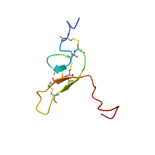
To analyze structural features of ω-Aga IVA, a gating modifier toxin from spider venom, we here investigated the NMR solution structure of ω-Aga IVA within DPC micelles. Under those conditions, the Cys-rich central region of ω-Aga IVA still retains the inhibitor Cys knot motif with three short antiparallel β-strands seen in water. However, 15 N HSQC spectra of ω-Aga IVA within micelles revealed that there are radical changes to the toxin's C-terminal tail and several loops upon binding to micelles. The C-terminal tail of ω-Aga IVA appears to assume a β-turn like conformation within micelles, though it is disordered in water. Whole-cell patch clamp studies with several ω-Aga IVA analogs indicate that both the hydrophobic C-terminal tail and an Arg patch in the core region of ω-Aga IVA are critical for Cav2.1 blockade. These results suggest that the membrane environment stabilizes the structure of the toxin, enabling it to act in a manner similar to other gating modifier toxins, though its mode of interaction with the membrane and the channel is unique.
- School of Life Sciences, Gwangju Institute of Science and Technology, 123, Cheomdangwagi-ro, Buk-gu, Gwangju, Republic of Korea.
Organizational Affiliation: