D19S Mutation of the Cationic, Cysteine-Rich Protein PAF: Novel Insights into Its Structural Dynamics, Thermal Unfolding and Antifungal Function.
Sonderegger, C., Fizil, A., Burtscher, L., Hajdu, D., Munoz, A., Gaspari, Z., Read, N.D., Batta, G., Marx, F.(2017) PLoS One 12: e0169920-e0169920
- PubMed: 28072824
- DOI: https://doi.org/10.1371/journal.pone.0169920
- Primary Citation of Related Structures:
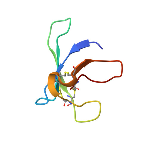
2NB0 - PubMed Abstract:
The cysteine-rich, cationic, antifungal protein PAF is abundantly secreted into the culture supernatant of the filamentous Ascomycete Penicillium chrysogenum. The five β-strands of PAF form a compact β-barrel that is stabilized by three disulphide bonds. The folding of PAF allows the formation of four surface-exposed loops and distinct charged motifs on the protein surface that might regulate the interaction of PAF with the sensitive target fungus. The growth inhibitory activity of this highly stable protein against opportunistic fungal pathogens provides great potential in antifungal drug research. To understand its mode of action, we started to investigate the surface-exposed loops of PAF and replaced one aspartic acid at position 19 in loop 2 that is potentially involved in PAF active or binding site, with a serine (Asp19 to Ser19). We analysed the overall effects, such as unfolding, electrostatic changes, sporadic conformers and antifungal activity when substituting this specific amino acid to the fairly indifferent amino acid serine. Structural analyses revealed that the overall 3D solution structure is virtually identical with that of PAF. However, PAFD19S showed slightly increased dynamics and significant differences in the surface charge distribution. Thermal unfolding identified PAFD19S to be rather a two-state folder in contrast to the three-state folder PAF. Functional comparison of PAFD19S and PAF revealed that the exchange at residue 19 caused a dramatic loss of antifungal activity: the binding and internalization of PAFD19S by target cells was reduced and the protein failed to trigger an intracellular Ca2+ response, all of which are closely linked to the antifungal toxicity of PAF. We conclude that the negatively charged residue Asp19 in loop 2 is essential for full function of the cationic protein PAF.
- Division of Molecular Biology, Biocenter, Medical University of Innsbruck, Innsbruck, Austria.
Organizational Affiliation: