Role of the Two Structural Domains from the Periplasmic Escherichia coli Histidine-binding Protein HisJ.
Chu, B.C., Dewolf, T., Vogel, H.J.(2013) J Biological Chem 288: 31409-31422
- PubMed: 24036119
- DOI: https://doi.org/10.1074/jbc.M113.490441
- Primary Citation of Related Structures:
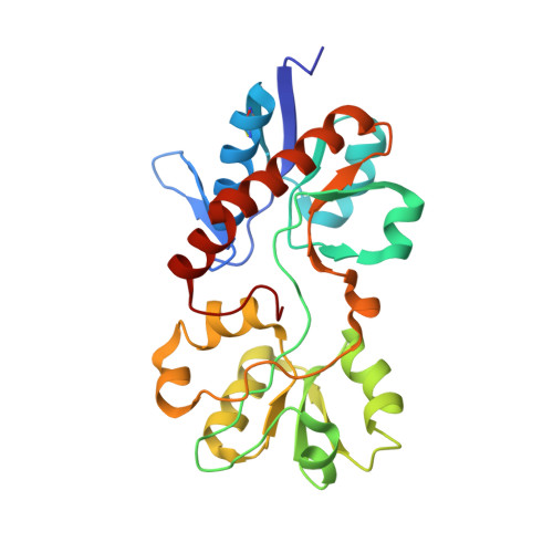
2M8C - PubMed Abstract:
Escherichia coli HisJ is a type II periplasmic binding protein that functions to reversibly capture histidine and transfer it to its cognate inner membrane ABC permease. Here, we used NMR spectroscopy to determine the structure of apo-HisJ (26.5 kDa) in solution. HisJ is a bilobal protein in which domain 1 (D1) is made up of two noncontiguous subdomains, and domain 2 (D2) is expressed as the inner domain. To better understand the roles of D1 and D2, we have isolated and characterized each domain separately. Structurally, D1 closely resembles its homologous domain in apo- and holo-HisJ, whereas D2 is more similar to the holo-form. NMR relaxation experiments reveal that HisJ becomes more ordered upon ligand binding, whereas isolated D2 experiences a significant reduction in slower (millisecond to microsecond) motions compared with the homologous domain in apo-HisJ. NMR titrations reveal that D1 is able to bind histidine in a similar manner as full-length HisJ, albeit with lower affinity. Unexpectedly, isolated D1 and D2 do not interact with each other in the presence or absence of histidine, which indicates the importance of intact interdomain-connecting elements (i.e. hinge regions) for HisJ functioning. Our results shed light on the binding mechanism of type II periplasmic binding proteins where ligand is initially bound by D1, and D2 plays a supporting role in this dynamic process.
- From the Biochemistry Research Group, Department of Biological Sciences, University of Calgary, Alberta T2N 1N4, Canada.
Organizational Affiliation: