2IZQ

Gramicidin D complex with KI


Experimental Data Snapshot

  • Method: X-RAY DIFFRACTION
  • Resolution: 0.80 Å
  • R-Value Free: 0.122 
  • R-Value Observed: 0.111 

wwPDB Validation   3D Report Full Report


This is version 4.1 of the entry. See complete history


Literature

Nonstoichiometric Complex of Gramicidin D with Ki at 0.80 A Resolution.

Olczak, A.Glowka, M.L.Szczesio, M.Bojarska, J.Duax, W.L.Burkhart, B.M.Wawrzak, Z.

(2007) Acta Crystallogr D Biol Crystallogr 63: 319

  • DOI: https://doi.org/10.1107/S0907444906053649
  • Primary Citation of Related Structures:  
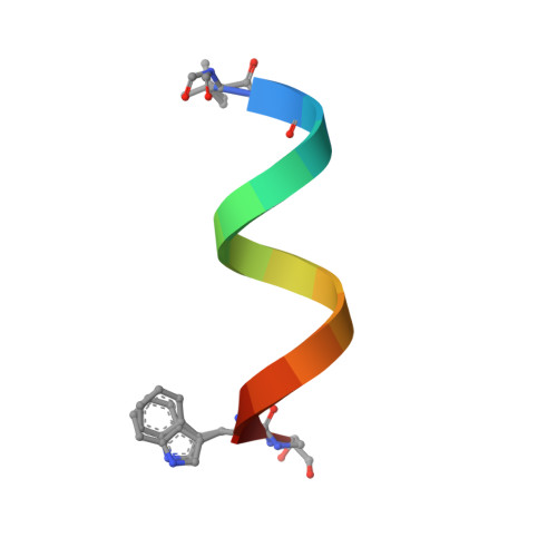
    2IZQ

  • PubMed Abstract: 

    The crystal structure of a nonstoichiometric complex of gramicidin D (gD) with KI has been determined at 100 K using synchrotron radiation. The final R factor was 0.106 for 83 988 observed reflections (Friedel pairs were not merged) collected to 0.80 A. The structure consists of four independent pentadecapeptides and numerous solvent molecules and salt ions. The general architecture of the antiparallel double-stranded gramicidin dimers in the crystal (a right-handed antiparallel DSbetaH(R) form) closely resembles that of previously published cation complexes of gD. However, a significantly different mixture of gramicidin isomers is found in the crystal of the KI complex, including partial occupancy of phenylalanine at position 11. Only three sites in each of the two crystallographically independent channels are partially occupied by potassium cations instead of the commonly observed seven sites. The sum of the partial occupancies of K(+) (1.10 per two dimers) is consistent with the sum of the iodide occupancies (1.095 over eight sites), which is also confirmed by the anomalous signal of the iodide. There was a significant asymmetry of the distribution and occupancies of cations in the crystallographically independent gramicidin channels, in contrast to the distribution found in the rubidium chloride complex with gD.


  • Organizational Affiliation

    Institute of General and Ecological Chemistry, University of Technology, Łódź, Poland.


Macromolecules

Find similar proteins by:  Sequence   |   3D Structure  

Entity ID: 1
MoleculeChains Sequence LengthOrganismDetailsImage
GRAMICIDIN D
A, B, C, D
16Brevibacillus brevisMutation(s): 2 
Entity Groups  
Sequence Clusters30% Identity50% Identity70% Identity90% Identity95% Identity100% Identity
Sequence Annotations
Expand
  • Reference Sequence
Small Molecules
Ligands 3 Unique
IDChains Name / Formula / InChI Key2D Diagram3D Interactions
IOD
Query on IOD

Download Ideal Coordinates CCD File 
G [auth A]
H [auth A]
I [auth A]
M [auth A]
N [auth B]
G [auth A],
H [auth A],
I [auth A],
M [auth A],
N [auth B],
O [auth B],
W [auth D],
X [auth D]
IODIDE ION
I
XMBWDFGMSWQBCA-UHFFFAOYSA-M
K
Query on K

Download Ideal Coordinates CCD File 
E [auth A]
F [auth A]
K [auth A]
T [auth C]
U [auth C]
E [auth A],
F [auth A],
K [auth A],
T [auth C],
U [auth C],
V [auth C]
POTASSIUM ION
K
NPYPAHLBTDXSSS-UHFFFAOYSA-N
MOH
Query on MOH

Download Ideal Coordinates CCD File 
J [auth A]
L [auth A]
P [auth C]
Q [auth C]
R [auth C]
J [auth A],
L [auth A],
P [auth C],
Q [auth C],
R [auth C],
S [auth C],
Y [auth D]
METHANOL
C H4 O
OKKJLVBELUTLKV-UHFFFAOYSA-N
Modified Residues  1 Unique
IDChains TypeFormula2D DiagramParent
FVA
Query on FVA
A, B, C, D
L-PEPTIDE LINKINGC6 H11 N O3VAL
Biologically Interesting Molecules (External Reference) 1 Unique
Experimental Data & Validation

Experimental Data

  • Method: X-RAY DIFFRACTION
  • Resolution: 0.80 Å
  • R-Value Free: 0.122 
  • R-Value Observed: 0.111 
  • Space Group: P 21 21 21
Unit Cell:
Length ( Å )Angle ( ˚ )
a = 30.144α = 90
b = 31.319β = 90
c = 51.554γ = 90
Software Package:
Software NamePurpose
SHELXL-97refinement
DENZOdata reduction
SCALEPACKdata scaling
AMoREphasing

Structure Validation

View Full Validation Report


Entry History 

Deposition Data

Revision History  (Full details and data files)

  • Version 1.0: 2007-01-23
    Type: Initial release
  • Version 1.1: 2011-07-13
    Changes: Atomic model, Database references, Derived calculations, Structure summary, Version format compliance
  • Version 1.2: 2011-07-20
    Changes: Derived calculations, Other, Structure summary
  • Version 1.3: 2012-11-30
    Changes: Other
  • Version 2.0: 2018-10-24
    Changes: Advisory, Atomic model, Data collection, Derived calculations
  • Version 3.0: 2019-01-23
    Changes: Atomic model, Data collection, Derived calculations
  • Version 3.1: 2019-05-22
    Changes: Data collection, Refinement description
  • Version 3.2: 2023-03-08
    Changes: Database references, Derived calculations, Other
  • Version 4.0: 2023-11-15
    Changes: Atomic model, Data collection, Derived calculations
  • Version 4.1: 2024-02-07
    Changes: Refinement description