2HEW

The X-ray crystal structure of murine OX40L


Experimental Data Snapshot

  • Method: X-RAY DIFFRACTION
  • Resolution: 1.45 Å
  • R-Value Free: 0.192 
  • R-Value Work: 0.165 
  • R-Value Observed: 0.168 

wwPDB Validation   3D Report Full Report


This is version 1.3 of the entry. See complete history


Literature

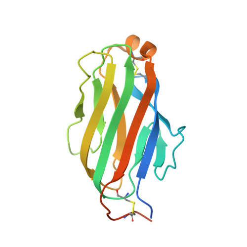
The Crystal Structure of the Costimulatory OX40-OX40L Complex.

Compaan, D.M.Hymowitz, S.G.

(2006) Structure 14: 1321-1330

  • DOI: https://doi.org/10.1016/j.str.2006.06.015
  • Primary Citation of Related Structures:  
    2HEV, 2HEW, 2HEY

  • PubMed Abstract: 

    OX40 is a T cell costimulator activated by OX40L. Blockade of the OX40L-OX40 interaction has ameliorative effects in animal models of T cell pathologies. In order to better understand the interaction between OX40 and OX40L, we have determined the crystal structure of murine OX40L and of the human OX40-OX40L complex at 1.45 and 2.4 A, respectively. These structures show that OX40L is an unusually small member of the tumor necrosis factor superfamily (TNFSF). The arrangement of the OX40L protomers forming the functional trimer is atypical and differs from that of other members by a 15 degrees rotation of each protomer with respect to the trimer axis, resulting in an open assembly. Site-directed changes of the interfacial residues of OX40L suggest this interface lacks a single "hot spot" and that instead, binding energy is dispersed over at least two distinct areas. These structures demonstrate the structural plasticity of TNFSF members and their interactions with receptors.


  • Organizational Affiliation

    Department of Protein Engineering, Genentech, Incorporated, 1 DNA Way, South San Francisco, California 94080, USA.


Macromolecules
Find similar proteins by:  (by identity cutoff)  |  3D Structure
Entity ID: 1
MoleculeChains Sequence LengthOrganismDetailsImage
Tumor necrosis factor ligand superfamily member 4A [auth F]152Mus musculusMutation(s): 0 
Gene Names: Tnfsf4Ox40lTxgp1l
Membrane Entity: Yes 
UniProt
Find proteins for P43488 (Mus musculus)
Explore P43488 
Go to UniProtKB:  P43488
Entity Groups  
Sequence Clusters30% Identity50% Identity70% Identity90% Identity95% Identity100% Identity
UniProt GroupP43488
Sequence Annotations
Expand
  • Reference Sequence
Small Molecules
Ligands 2 Unique
IDChains Name / Formula / InChI Key2D Diagram3D Interactions
NAG
Query on NAG

Download Ideal Coordinates CCD File 
B [auth F]2-acetamido-2-deoxy-beta-D-glucopyranose
C8 H15 N O6
OVRNDRQMDRJTHS-FMDGEEDCSA-N
SO4
Query on SO4

Download Ideal Coordinates CCD File 
C [auth F]SULFATE ION
O4 S
QAOWNCQODCNURD-UHFFFAOYSA-L
Experimental Data & Validation

Experimental Data

  • Method: X-RAY DIFFRACTION
  • Resolution: 1.45 Å
  • R-Value Free: 0.192 
  • R-Value Work: 0.165 
  • R-Value Observed: 0.168 
  • Space Group: P 63
Unit Cell:
Length ( Å )Angle ( ˚ )
a = 74.149α = 90
b = 74.149β = 90
c = 48.815γ = 120
Software Package:
Software NamePurpose
REFMACrefinement
SHARPphasing

Structure Validation

View Full Validation Report


Entry History 

Deposition Data

Revision History  (Full details and data files)

  • Version 1.0: 2006-08-29
    Type: Initial release
  • Version 1.1: 2008-05-01
    Changes: Version format compliance
  • Version 1.2: 2011-07-13
    Changes: Derived calculations, Version format compliance
  • Version 1.3: 2020-07-29
    Type: Remediation
    Reason: Carbohydrate remediation
    Changes: Advisory, Data collection, Database references, Derived calculations, Structure summary