2HAP

STRUCTURE OF A HAP1-18/DNA COMPLEX REVEALS THAT PROTEIN/DNA INTERACTIONS CAN HAVE DIRECT ALLOSTERIC EFFECTS ON TRANSCRIPTIONAL ACTIVATION


Experimental Data Snapshot

  • Method: X-RAY DIFFRACTION
  • Resolution: 2.50 Å
  • R-Value Free: 0.302 
  • R-Value Work: 0.248 
  • R-Value Observed: 0.248 

wwPDB Validation   3D Report Full Report


This is version 1.4 of the entry. See complete history


Literature

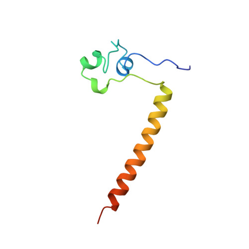
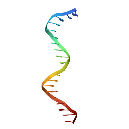
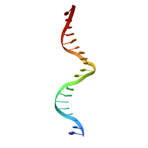
Structure of HAP1-18-DNA implicates direct allosteric effect of protein-DNA interactions on transcriptional activation.

King, D.A.Zhang, L.Guarente, L.Marmorstein, R.

(1999) Nat Struct Biol 6: 22-27

  • DOI: https://doi.org/10.1038/4893
  • Primary Citation of Related Structures:  
    2HAP

  • PubMed Abstract: 

    HAP1 is a yeast transcriptional activator that binds with equal affinity to the dissimilar upstream activation sequences UAS1 and UAS(CYC7), but activates transcription differentially when bound to each site. HAP1-18 harbors an amino acid change in the DNA binding domain. While binding UAS1 poorly, HAP1-18 binds UAS(CYC7) with wild-type properties and activates transcription at elevated levels relative to HAP1. We have determined the structure of HAP1-18-UAS(CYC7) and have compared it to HAP1-UAS(CYC7). Unexpectedly, the single amino acid substitution in HAP1-18 nucleates a significantly altered hydrogen bond interface between the protein and DNA resulting in DNA conformational changes and an ordering of one N-terminal arm of the protein dimer along the DNA minor groove. These observations, together with a large subset of transcriptionally defective mutations in the HAP1 DNA-binding domain that map to the HAP1-DNA interface, suggest that protein-DNA interactions may have direct allosteric effects on transcriptional activation.


  • Organizational Affiliation

    The Wistar Institute and The Department of Chemistry, University of Pennsylvania, Philadelphia 19104, USA.


Macromolecules

Find similar proteins by:  (by identity cutoff)  |  3D Structure
Entity ID: 3
MoleculeChains Sequence LengthOrganismDetailsImage
PROTEIN (HEME ACTIVATOR PROTEIN)
C, D
81Saccharomyces cerevisiaeMutation(s): 0 
UniProt
Find proteins for P0CS82 (Saccharomyces cerevisiae)
Explore P0CS82 
Go to UniProtKB:  P0CS82
Entity Groups  
Sequence Clusters30% Identity50% Identity70% Identity90% Identity95% Identity100% Identity
UniProt GroupP0CS82
Sequence Annotations
Expand
  • Reference Sequence

Find similar nucleic acids by:  Sequence   |   3D Structure  

Entity ID: 1
MoleculeChains LengthOrganismImage
DNA (5'-D(*AP*CP*GP*CP*TP*AP*TP*TP*AP*TP*CP*GP*CP*TP*AP*TP*TP*AP*GP*T)-3')20N/A
Sequence Annotations
Expand
  • Reference Sequence

Find similar nucleic acids by:  Sequence   |   3D Structure  

Entity ID: 2
MoleculeChains LengthOrganismImage
DNA (5'-D(*AP*CP*TP*AP*AP*TP*AP*GP*CP*GP*AP*TP*AP*AP*TP*AP*GP*CP*GP*T)-3')20N/A
Sequence Annotations
Expand
  • Reference Sequence
Experimental Data & Validation

Experimental Data

  • Method: X-RAY DIFFRACTION
  • Resolution: 2.50 Å
  • R-Value Free: 0.302 
  • R-Value Work: 0.248 
  • R-Value Observed: 0.248 
  • Space Group: P 41 21 2
Unit Cell:
Length ( Å )Angle ( ˚ )
a = 92.5α = 90
b = 92.5β = 90
c = 86.5γ = 90
Software Package:
Software NamePurpose
PHASESphasing
CCP4model building
X-PLORrefinement
XDSdata reduction
XSCALEdata scaling
CCP4phasing

Structure Validation

View Full Validation Report


Entry History 

Deposition Data

Revision History  (Full details and data files)

  • Version 1.0: 1999-11-10
    Type: Initial release
  • Version 1.1: 2008-05-22
    Changes: Version format compliance
  • Version 1.2: 2011-07-13
    Changes: Version format compliance
  • Version 1.3: 2011-11-16
    Changes: Atomic model
  • Version 1.4: 2024-02-14
    Changes: Data collection, Database references, Derived calculations