2GZQ

Phosphatidylethanolamine-binding protein from Plasmodium vivax


Experimental Data Snapshot

  • Method: X-RAY DIFFRACTION
  • Resolution: 1.30 Å
  • R-Value Free: 0.180 
  • R-Value Work: 0.161 

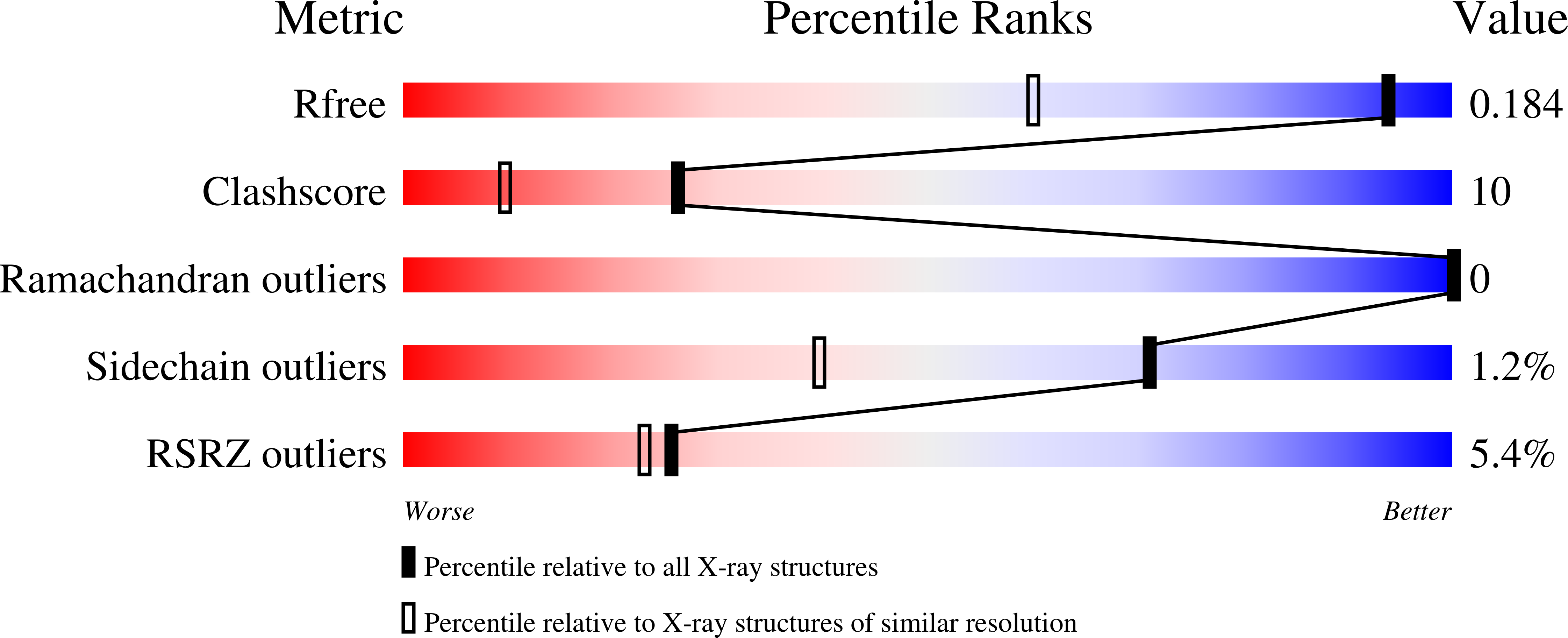
wwPDB Validation   3D Report Full Report


This is version 1.4 of the entry. See complete history


Literature

The structure of Plasmodium vivax phosphatidylethanolamine-binding protein suggests a functional motif containing a left-handed helix

Arakaki, T.Neely, H.Boni, E.Mueller, N.Buckner, F.S.Van Voorhis, W.C.Lauricella, A.DeTitta, G.Luft, J.Hol, W.G.Merritt, E.A.

(2007) Acta Crystallogr Sect F Struct Biol Cryst Commun 63: 178-182

  • DOI: https://doi.org/10.1107/S1744309107007580
  • Primary Citation of Related Structures:  
    2GZQ

  • PubMed Abstract: 

    The structure of a putative Raf kinase inhibitor protein (RKIP) homolog from the eukaryotic parasite Plasmodium vivax has been studied to a resolution of 1.3 A using multiple-wavelength anomalous diffraction at the Se K edge. This protozoan protein is topologically similar to previously studied members of the phosphatidylethanolamine-binding protein (PEBP) sequence family, but exhibits a distinctive left-handed alpha-helical region at one side of the canonical phospholipid-binding site. Re-examination of previously determined PEBP structures suggests that the P. vivax protein and yeast carboxypeptidase Y inhibitor may represent a structurally distinct subfamily of the diverse PEBP-sequence family.


  • Organizational Affiliation

    Structural Genomics of Pathogenic Protozoa Consortium, USA.


Macromolecules
Find similar proteins by:  (by identity cutoff)  |  3D Structure
Entity ID: 1
MoleculeChains Sequence LengthOrganismDetailsImage
Phosphatidylethanolamine-binding protein200Plasmodium vivaxMutation(s): 0 
Gene Names: hPf.PFL0955c
UniProt
Find proteins for A5K000 (Plasmodium vivax (strain Salvador I))
Explore A5K000 
Go to UniProtKB:  A5K000
Entity Groups  
Sequence Clusters30% Identity50% Identity70% Identity90% Identity95% Identity100% Identity
UniProt GroupA5K000
Sequence Annotations
Expand
  • Reference Sequence
Small Molecules
Modified Residues  1 Unique
IDChains TypeFormula2D DiagramParent
MSE
Query on MSE
A
L-PEPTIDE LINKINGC5 H11 N O2 SeMET
Experimental Data & Validation

Experimental Data

  • Method: X-RAY DIFFRACTION
  • Resolution: 1.30 Å
  • R-Value Free: 0.180 
  • R-Value Work: 0.161 
  • Space Group: P 21 21 21
Unit Cell:
Length ( Å )Angle ( ˚ )
a = 39.309α = 90
b = 54.11β = 90
c = 94.377γ = 90
Software Package:
Software NamePurpose
DENZOdata reduction
SCALEPACKdata scaling
SOLVEphasing
RESOLVEphasing
REFMACrefinement
PDB_EXTRACTdata extraction

Structure Validation

View Full Validation Report


Entry History 

Deposition Data

Revision History  (Full details and data files)

  • Version 1.0: 2006-05-23
    Type: Initial release
  • Version 1.1: 2008-05-01
    Changes: Version format compliance
  • Version 1.2: 2011-07-13
    Changes: Advisory, Version format compliance
  • Version 1.3: 2017-10-18
    Changes: Refinement description
  • Version 1.4: 2024-04-03
    Changes: Data collection, Database references, Derived calculations, Refinement description