


Crystal Structure of glucose-1-phosphate thymidylyltransferase from Sulfolobus tokodaii
Rajakannan, V., Yamane, T.To be published.
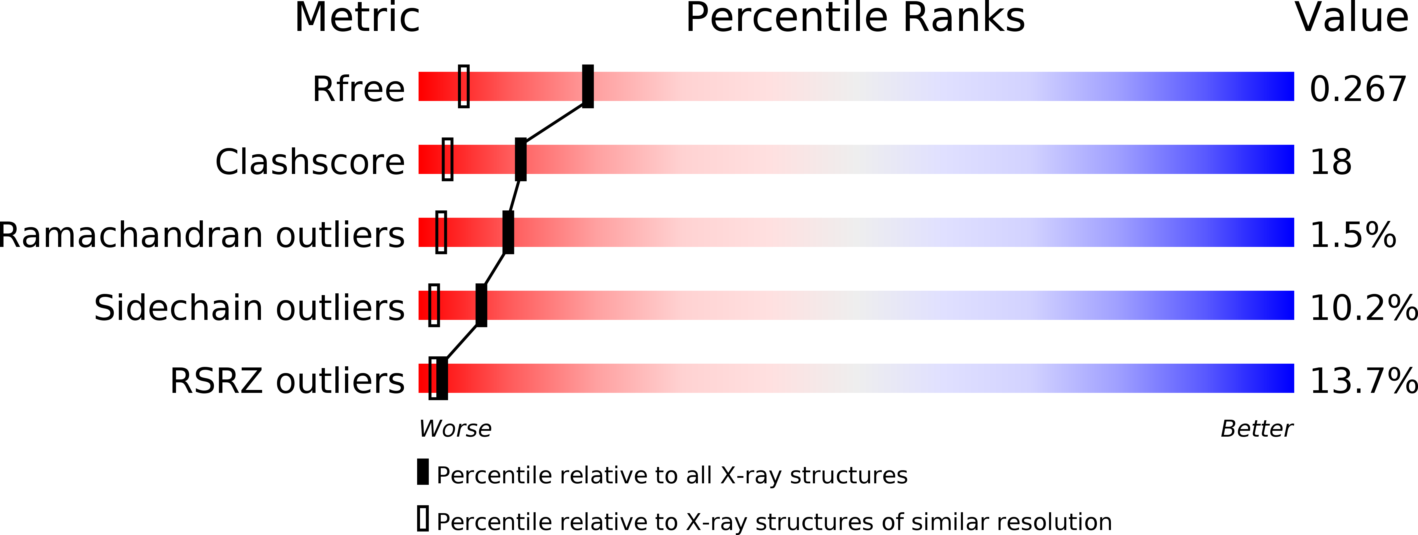
Experimental Data Snapshot
wwPDB Validation 3D Report Full Report
Entity ID: 1 | |||||
|---|---|---|---|---|---|
| Molecule | Chains | Sequence Length | Organism | Details | Image |
| 401aa long hypothetical glucose-1-phosphate thymidylyltransferase | 401 | Sulfurisphaera tokodaii | Mutation(s): 0 EC: 2.7.7.24 | 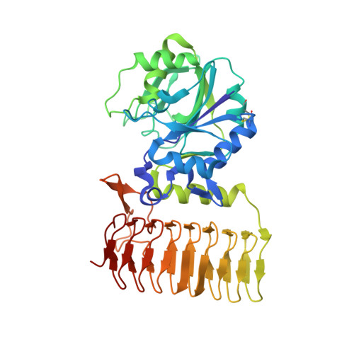 | |
UniProt | |||||
Find proteins for Q975F9 (Sulfurisphaera tokodaii (strain DSM 16993 / JCM 10545 / NBRC 100140 / 7)) Explore Q975F9 Go to UniProtKB: Q975F9 | |||||
Entity Groups | |||||
| Sequence Clusters | 30% Identity50% Identity70% Identity90% Identity95% Identity100% Identity | ||||
| UniProt Group | Q975F9 | ||||
Sequence AnnotationsExpand | |||||
| |||||
| Length ( Å ) | Angle ( ˚ ) |
|---|---|
| a = 122.999 | α = 90 |
| b = 122.999 | β = 90 |
| c = 94.884 | γ = 120 |
| Software Name | Purpose |
|---|---|
| REFMAC | refinement |
| CrystalClear | data reduction |
| SCALEPACK | data scaling |
| SHELXD | phasing |
RCSB PDB (citation) is hosted by
RCSB PDB is a member of the

