2EWJ

Escherichia Coli Replication Terminator Protein (Tus) Complexed With DNA- Locked form


Experimental Data Snapshot

  • Method: X-RAY DIFFRACTION
  • Resolution: 2.70 Å
  • R-Value Free: 0.303 
  • R-Value Work: 0.219 
  • R-Value Observed: 0.223 

wwPDB Validation   3D Report Full Report


This is version 1.3 of the entry. See complete history


Literature

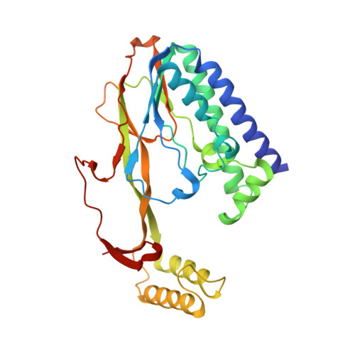
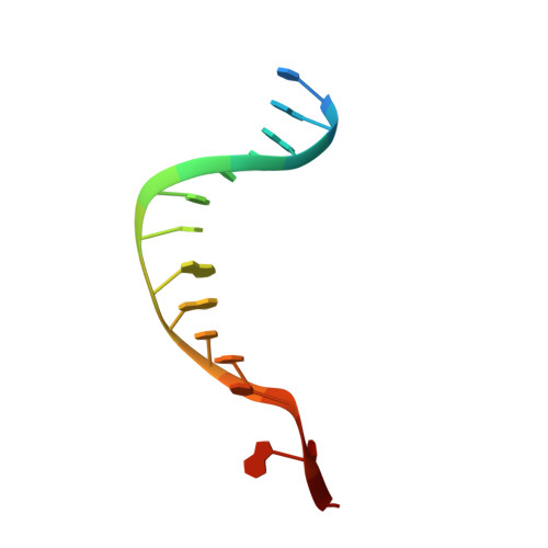
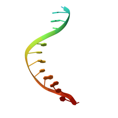
A molecular mousetrap determines polarity of termination of DNA replication in E. coli.

Mulcair, M.D.Schaeffer, P.M.Oakley, A.J.Cross, H.F.Neylon, C.Hill, T.M.Dixon, N.E.

(2006) Cell 125: 1309-1319

  • DOI: https://doi.org/10.1016/j.cell.2006.04.040
  • Primary Citation of Related Structures:  
    2EWJ

  • PubMed Abstract: 

    During chromosome synthesis in Escherichia coli, replication forks are blocked by Tus bound Ter sites on approach from one direction but not the other. To study the basis of this polarity, we measured the rates of dissociation of Tus from forked TerB oligonucleotides, such as would be produced by the replicative DnaB helicase at both the fork-blocking (nonpermissive) and permissive ends of the Ter site. Strand separation of a few nucleotides at the permissive end was sufficient to force rapid dissociation of Tus to allow fork progression. In contrast, strand separation extending to and including the strictly conserved G-C(6) base pair at the nonpermissive end led to formation of a stable locked complex. Lock formation specifically requires the cytosine residue, C(6). The crystal structure of the locked complex showed that C(6) moves 14 A from its normal position to bind in a cytosine-specific pocket on the surface of Tus.


  • Organizational Affiliation

    Research School of Chemistry, Australian National University, Canberra, ACT 0200, Australia.


Macromolecules

Find similar proteins by:  (by identity cutoff)  |  3D Structure
Entity ID: 3
MoleculeChains Sequence LengthOrganismDetailsImage
DNA replication terminus site-binding proteinC [auth A]309Escherichia coliMutation(s): 0 
Gene Names: tus
UniProt
Find proteins for P16525 (Escherichia coli (strain K12))
Explore P16525 
Go to UniProtKB:  P16525
Entity Groups  
Sequence Clusters30% Identity50% Identity70% Identity90% Identity95% Identity100% Identity
UniProt GroupP16525
Sequence Annotations
Expand
  • Reference Sequence

Find similar nucleic acids by:  Sequence   |   3D Structure  

Entity ID: 1
MoleculeChains LengthOrganismImage
5'-D(*T*TP*AP*GP*TP*TP*AP*CP*AP*AP*CP*AP*TP*AP*CP*T)-3'A [auth B]16N/A
Sequence Annotations
Expand
  • Reference Sequence

Find similar nucleic acids by:  Sequence   |   3D Structure  

Entity ID: 2
MoleculeChains LengthOrganismImage
5'-D(*TP*G*AP*TP*AP*TP*GP*TP*TP*GP*TP*AP*AP*CP*TP*A)-3'B [auth C]16N/A
Sequence Annotations
Expand
  • Reference Sequence
Experimental Data & Validation

Experimental Data

  • Method: X-RAY DIFFRACTION
  • Resolution: 2.70 Å
  • R-Value Free: 0.303 
  • R-Value Work: 0.219 
  • R-Value Observed: 0.223 
  • Space Group: P 41 21 2
Unit Cell:
Length ( Å )Angle ( ˚ )
a = 62.679α = 90
b = 62.679β = 90
c = 251.619γ = 90
Software Package:
Software NamePurpose
REFMACrefinement
DENZOdata reduction
SCALEPACKdata scaling
MOLREPphasing

Structure Validation

View Full Validation Report


Entry History 

Deposition Data

Revision History  (Full details and data files)

  • Version 1.0: 2006-05-03
    Type: Initial release
  • Version 1.1: 2008-05-01
    Changes: Version format compliance
  • Version 1.2: 2011-07-13
    Changes: Advisory, Version format compliance
  • Version 1.3: 2023-10-25
    Changes: Data collection, Database references, Derived calculations, Refinement description