2EAX

Crystal structure of human PGRP-IBETAC in complex with glycosamyl muramyl pentapeptide


Experimental Data Snapshot

  • Method: X-RAY DIFFRACTION
  • Resolution: 2.10 Å
  • R-Value Free: 0.242 
  • R-Value Work: 0.210 
  • R-Value Observed: 0.215 

wwPDB Validation   3D Report Full Report


This is version 2.1 of the entry. See complete history


Literature

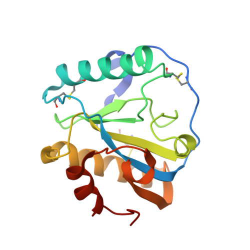
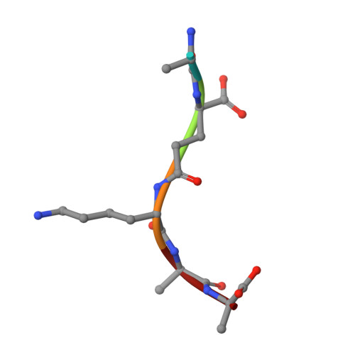
Structural insights into the bactericidal mechanism of human peptidoglycan recognition proteins

Cho, S.Wang, Q.Swaminathan, C.P.Hesek, D.Lee, M.Boons, G.J.Mobashery, S.Mariuzza, R.A.

(2007) Proc Natl Acad Sci U S A 104: 8761-8766

  • DOI: https://doi.org/10.1073/pnas.0701453104
  • Primary Citation of Related Structures:  
    2EAV, 2EAX

  • PubMed Abstract: 

    Peptidoglycan recognition proteins (PGRPs) are highly conserved pattern-recognition molecules of the innate immune system that bind bacterial peptidoglycans (PGNs), which are polymers of alternating N-acetylglucosamine (NAG) and N-acetylmuramic acid (NAM) cross-linked by short peptide stems. Human PRGPs are bactericidal against pathogenic and nonpathogenic Gram-positive bacteria, but not normal flora bacteria. Like certain glycopeptide antibiotics (e.g., vancomycin), PGRPs kill bacteria by directly interacting with their cell wall PGN, thereby interfering with PGN maturation. To better understand the bactericidal mechanism of PGRPs, we determined the crystal structure of the C-terminal PGN-binding domain of human PGRP-I beta in complex with NAG-NAM-L-Ala-gamma-D-Glu-L-Lys-D-Ala-D-Ala, a synthetic glycopeptide comprising a complete PGN repeat. This structure, in conjunction with the previously reported NMR structure of a dimeric PGN fragment, permitted identification of major conformational differences between free and PGRP-bound PGN with respect to the relative orientation of saccharide and peptide moieties. These differences provided structural insights into the bactericidal mechanism of human PGRPs. On the basis of molecular modeling, we propose that these proteins disrupt cell wall maturation not only by sterically encumbering access of biosynthetic enzymes to the nascent PGN chains, but also by locking PGN into a conformation that prevents formation of cross-links between peptide stems in the growing cell wall.


  • Organizational Affiliation

    Center for Advanced Research in Biotechnology, W. M. Keck Laboratory for Structural Biology, University of Maryland Biotechnology Institute, 9600 Gudelsky Drive, Rockville, MD 20850, USA.


Macromolecules
Find similar proteins by:  (by identity cutoff)  |  3D Structure
Entity ID: 1
MoleculeChains Sequence LengthOrganismDetailsImage
Peptidoglycan recognition protein-I-beta
A, B, C
164Homo sapiensMutation(s): 0 
Gene Names: PGRPIB
UniProt & NIH Common Fund Data Resources
Find proteins for Q96LB8 (Homo sapiens)
Explore Q96LB8 
Go to UniProtKB:  Q96LB8
PHAROS:  Q96LB8
GTEx:  ENSG00000163218 
Entity Groups  
Sequence Clusters30% Identity50% Identity70% Identity90% Identity95% Identity100% Identity
UniProt GroupQ96LB8
Sequence Annotations
Expand
  • Reference Sequence

Find similar proteins by:  Sequence   |   3D Structure  

Entity ID: 2
MoleculeChains Sequence LengthOrganismDetailsImage
GLYCOSAMYL MURAMYL PENTAPEPTIDED [auth L]5Homo sapiensMutation(s): 0 
Sequence Annotations
Expand
  • Reference Sequence
Oligosaccharides

Help

Entity ID: 3
MoleculeChains Length2D Diagram Glycosylation3D Interactions
2-acetamido-2-deoxy-beta-D-glucopyranose-(1-4)-methyl 2-acetamido-3-O-[(1R)-1-carboxyethyl]-2-deoxy-beta-D-glucopyranosideE [auth D]2N/A
Glycosylation Resources
GlyTouCan:  G69619HL
GlyCosmos:  G69619HL
Experimental Data & Validation

Experimental Data

  • Method: X-RAY DIFFRACTION
  • Resolution: 2.10 Å
  • R-Value Free: 0.242 
  • R-Value Work: 0.210 
  • R-Value Observed: 0.215 
  • Space Group: P 1 21 1
Unit Cell:
Length ( Å )Angle ( ˚ )
a = 41.94α = 90
b = 61.39β = 96.45
c = 97.95γ = 90
Software Package:
Software NamePurpose
CrystalCleardata collection
MOLREPphasing
CNSrefinement
CrystalCleardata reduction
CrystalCleardata scaling

Structure Validation

View Full Validation Report


Entry History 

Deposition Data

  • Released Date: 2007-10-02 
  • Deposition Author(s): Cho, S.

Revision History  (Full details and data files)

  • Version 1.0: 2007-10-02
    Type: Initial release
  • Version 1.1: 2011-07-13
    Changes: Version format compliance
  • Version 1.2: 2011-07-27
    Changes: Database references, Derived calculations, Non-polymer description
  • Version 1.3: 2011-09-14
    Changes: Non-polymer description
  • Version 1.4: 2011-09-21
    Changes: Source and taxonomy, Structure summary
  • Version 1.5: 2014-12-17
    Changes: Atomic model, Derived calculations
  • Version 2.0: 2020-07-29
    Type: Remediation
    Reason: Carbohydrate remediation
    Changes: Advisory, Atomic model, Data collection, Derived calculations, Structure summary
  • Version 2.1: 2023-10-25
    Changes: Data collection, Database references, Refinement description, Structure summary