


Crystal Structure of Hypothetical protein from Aeropyrum pernix
Lokanath, N.K., Kunishima, N.To be published.
Experimental Data Snapshot
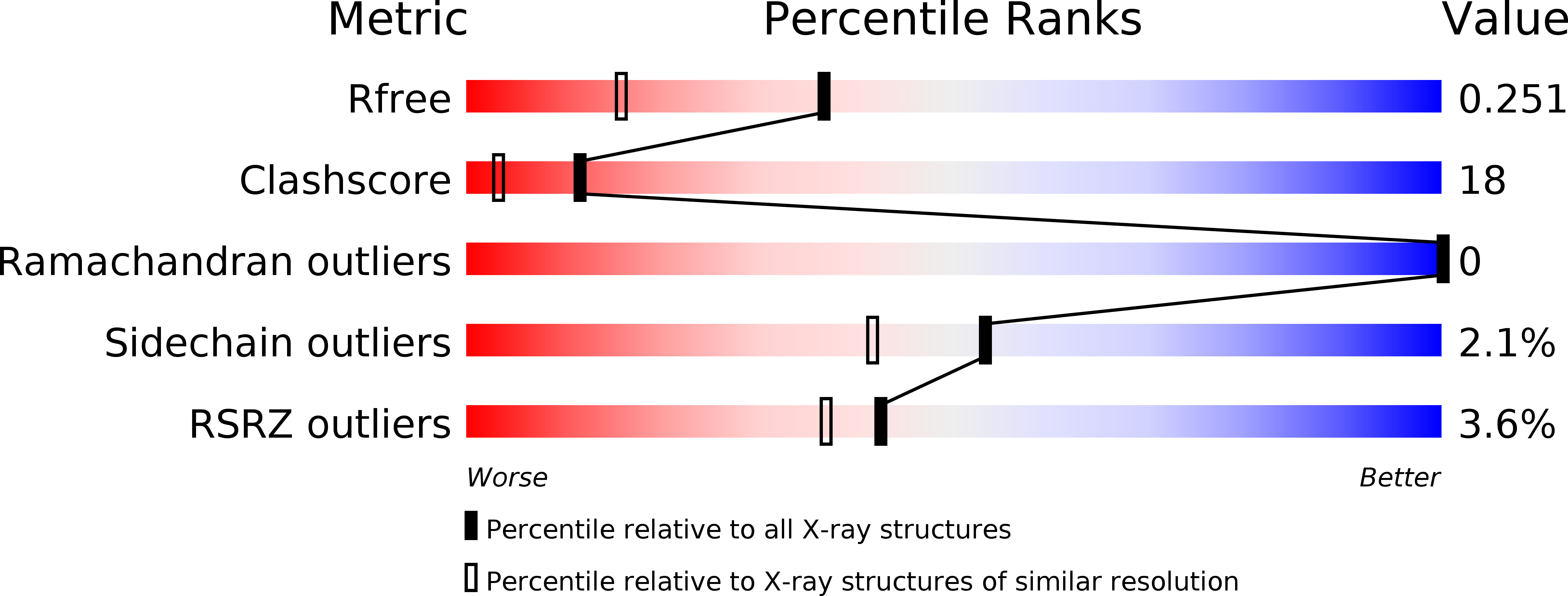
wwPDB Validation 3D Report Full Report
Entity ID: 1 | |||||
|---|---|---|---|---|---|
| Molecule | Chains | Sequence Length | Organism | Details | Image |
| UPF0130 protein APE0816 | 188 | Aeropyrum pernix | Mutation(s): 0 | 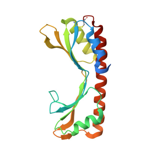 | |
UniProt | |||||
Find proteins for Q9YDV3 (Aeropyrum pernix (strain ATCC 700893 / DSM 11879 / JCM 9820 / NBRC 100138 / K1)) Explore Q9YDV3 Go to UniProtKB: Q9YDV3 | |||||
Entity Groups | |||||
| Sequence Clusters | 30% Identity50% Identity70% Identity90% Identity95% Identity100% Identity | ||||
| UniProt Group | Q9YDV3 | ||||
Sequence AnnotationsExpand | |||||
| |||||
| Length ( Å ) | Angle ( ˚ ) |
|---|---|
| a = 55.87 | α = 90 |
| b = 70.232 | β = 90 |
| c = 91.579 | γ = 90 |
| Software Name | Purpose |
|---|---|
| CNS | refinement |
| HKL-2000 | data reduction |
| SCALEPACK | data scaling |
| SOLVE | phasing |
RCSB PDB (citation) is hosted by
RCSB PDB is a member of the

