2D7F

Crystal structure of A lectin from canavalia gladiata seeds complexed with alpha-methyl-mannoside and alpha-aminobutyric acid


Experimental Data Snapshot

  • Method: X-RAY DIFFRACTION
  • Resolution: 2.31 Å
  • R-Value Free: 0.227 
  • R-Value Work: 0.180 
  • R-Value Observed: 0.182 

wwPDB Validation   3D Report Full Report


Ligand Structure Quality Assessment 


This is version 1.4 of the entry. See complete history


Literature

Structure of a lectin from Canavalia gladiata seeds: new structural insights for old molecules

Delatorre, P.Rocha, B.A.M.Souza, E.P.Oliveira, T.M.Bezerra, G.A.Moreno, F.B.M.B.Freitas, B.T.Santi-Gadelha, T.Sampaio, A.H.Azevedo Jr., W.F.Cavada, B.S.

(2007) BMC Struct Biol 7: 52-52

  • DOI: https://doi.org/10.1186/1472-6807-7-52
  • Primary Citation of Related Structures:  
    1WUV, 2D7F

  • PubMed Abstract: 

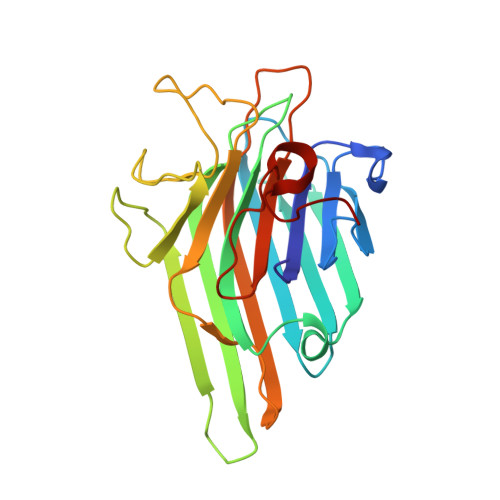
    Lectins are mainly described as simple carbohydrate-binding proteins. Previous studies have tried to identify other binding sites, which possible recognize plant hormones, secondary metabolites, and isolated amino acid residues. We report the crystal structure of a lectin isolated from Canavalia gladiata seeds (CGL), describing a new binding pocket, which may be related to pathogen resistance activity in ConA-like lectins; a site where a non-protein amino-acid, alpha-aminobutyric acid (Abu), is bound. The overall structure of native CGL and complexed with alpha-methyl-mannoside and Abu have been refined at 2.3 A and 2.31 A resolution, respectively. Analysis of the electron density maps of the CGL structure shows clearly the presence of Abu, which was confirmed by mass spectrometry. The presence of Abu in a plant lectin structure strongly indicates the ability of lectins on carrying secondary metabolites. Comparison of the amino acids composing the site with other legume lectins revealed that this site is conserved, providing an evidence of the biological relevance of this site. This new action of lectins strengthens their role in defense mechanisms in plants.


  • Organizational Affiliation

    Departamento de Bioquímica e Biologia Molecular, Universidade Federal do Ceará, Ceará, Brazil. plinio@urca.br


Macromolecules
Find similar proteins by:  (by identity cutoff)  |  3D Structure
Entity ID: 1
MoleculeChains Sequence LengthOrganismDetailsImage
Concanavalin AA,
B [auth F],
C [auth L],
D [auth S]
237Canavalia gladiataMutation(s): 0 
UniProt
Find proteins for P14894 (Canavalia gladiata)
Explore P14894 
Go to UniProtKB:  P14894
Entity Groups  
Sequence Clusters30% Identity50% Identity70% Identity90% Identity95% Identity100% Identity
UniProt GroupP14894
Sequence Annotations
Expand
  • Reference Sequence
Small Molecules
Ligands 4 Unique
IDChains Name / Formula / InChI Key2D Diagram3D Interactions
MMA
Query on MMA

Download Ideal Coordinates CCD File 
E [auth A],
I [auth F],
M [auth L],
Q [auth S]
methyl alpha-D-mannopyranoside
C7 H14 O6
HOVAGTYPODGVJG-VEIUFWFVSA-N
DBB
Query on DBB

Download Ideal Coordinates CCD File 
H [auth A],
L [auth F],
P [auth L],
T [auth S]
D-ALPHA-AMINOBUTYRIC ACID
C4 H9 N O2
QWCKQJZIFLGMSD-GSVOUGTGSA-N
MN
Query on MN

Download Ideal Coordinates CCD File 
F [auth A],
J [auth F],
N [auth L],
R [auth S]
MANGANESE (II) ION
Mn
WAEMQWOKJMHJLA-UHFFFAOYSA-N
CA
Query on CA

Download Ideal Coordinates CCD File 
G [auth A],
K [auth F],
O [auth L],
S
CALCIUM ION
Ca
BHPQYMZQTOCNFJ-UHFFFAOYSA-N
Experimental Data & Validation

Experimental Data

  • Method: X-RAY DIFFRACTION
  • Resolution: 2.31 Å
  • R-Value Free: 0.227 
  • R-Value Work: 0.180 
  • R-Value Observed: 0.182 
  • Space Group: C 2 2 21
Unit Cell:
Length ( Å )Angle ( ˚ )
a = 100.915α = 90
b = 115.754β = 90
c = 241.626γ = 90
Software Package:
Software NamePurpose
REFMACrefinement
MOSFLMdata reduction
CCP4data scaling
MOLREPphasing

Structure Validation

View Full Validation Report


Ligand Structure Quality Assessment 


Entry History 

Deposition Data

Revision History  (Full details and data files)

  • Version 1.0: 2006-12-19
    Type: Initial release
  • Version 1.1: 2007-11-21
    Changes: Version format compliance
  • Version 1.2: 2011-07-13
    Changes: Version format compliance
  • Version 1.3: 2020-07-29
    Type: Remediation
    Reason: Carbohydrate remediation
    Changes: Advisory, Data collection, Database references, Derived calculations, Source and taxonomy, Structure summary
  • Version 1.4: 2024-03-13
    Changes: Data collection, Database references, Structure summary