2C2V

Crystal structure of the CHIP-UBC13-UEV1a complex


Experimental Data Snapshot

  • Method: X-RAY DIFFRACTION
  • Resolution: 2.90 Å
  • R-Value Free: 0.297 
  • R-Value Work: 0.214 
  • R-Value Observed: 0.218 

wwPDB Validation   3D Report Full Report


This is version 1.5 of the entry. See complete history


Literature

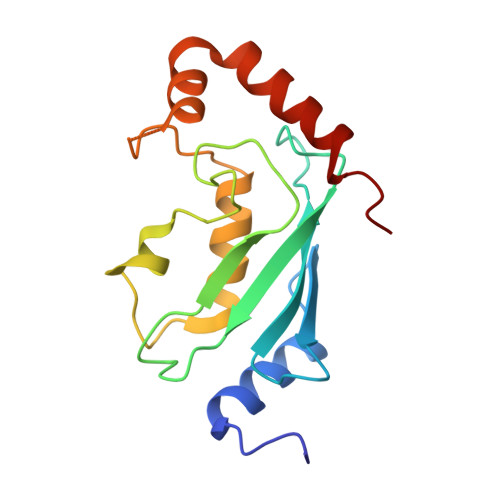
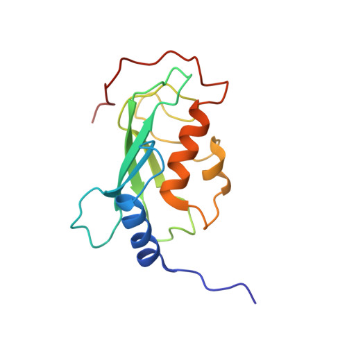
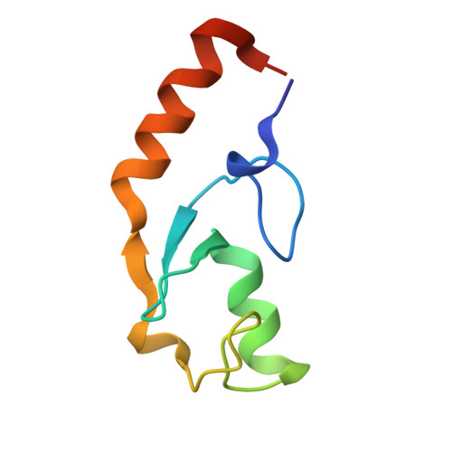
Chaperoned ubiquitylation--crystal structures of the CHIP U box E3 ubiquitin ligase and a CHIP-Ubc13-Uev1a complex.

Zhang, M.Windheim, M.Roe, S.M.Peggie, M.Cohen, P.Prodromou, C.Pearl, L.H.

(2005) Mol Cell 20: 525-538

  • DOI: https://doi.org/10.1016/j.molcel.2005.09.023
  • Primary Citation of Related Structures:  
    2C2L, 2C2V

  • PubMed Abstract: 

    CHIP is a dimeric U box E3 ubiquitin ligase that binds Hsp90 and/or Hsp70 via its TPR-domain, facilitating ubiquitylation of chaperone bound client proteins. We have determined the crystal structure of CHIP bound to an Hsp90 C-terminal decapeptide. The structure explains how CHIP associates with either chaperone type and reveals an unusual asymmetric homodimer in which the protomers adopt radically different conformations. Additionally, we identified CHIP as a functional partner of Ubc13-Uev1a in formation of Lys63-linked polyubiquitin chains, extending CHIP's roles into ubiquitin regulation as well as targeted destruction. The structure of Ubc13-Uev1a bound to the CHIP U box domain defines the basis for selective cooperation of CHIP with specific ubiquitin-conjugating enzymes. Remarkably, the asymmetric arrangement of the TPR domains in the CHIP dimer occludes one Ubc binding site, so that CHIP operates with half-of-sites activity, providing an elegant means for coupling a dimeric chaperone to a single ubiquitylation system.


  • Organizational Affiliation

    Section of Structural Biology, Institute of Cancer Research, Chester Beatty Laboratories, 237 Fulham Road, London SW3 6JB, United Kingdom.


Macromolecules
Find similar proteins by:  (by identity cutoff)  |  3D Structure
Entity ID: 1
MoleculeChains Sequence LengthOrganismDetailsImage
Ubiquitin-conjugating enzyme E2 NA [auth B],
C [auth E],
E [auth H],
G [auth K]
154Homo sapiensMutation(s): 0 
Gene Names: UBE2NBLU
EC: 2.3.2.23
UniProt & NIH Common Fund Data Resources
Find proteins for P61088 (Homo sapiens)
Explore P61088 
Go to UniProtKB:  P61088
PHAROS:  P61088
GTEx:  ENSG00000177889 
Entity Groups  
Sequence Clusters30% Identity50% Identity70% Identity90% Identity95% Identity100% Identity
UniProt GroupP61088
Sequence Annotations
Expand
  • Reference Sequence
Find similar proteins by:  (by identity cutoff)  |  3D Structure
Entity ID: 2
MoleculeChains Sequence LengthOrganismDetailsImage
Ubiquitin-conjugating enzyme E2 variant 1B [auth C],
D [auth F],
F [auth I],
H [auth L]
142Homo sapiensMutation(s): 0 
Gene Names: UBE2V1CROC1UBE2VUEV1P/OKcl.19
UniProt & NIH Common Fund Data Resources
Find proteins for Q13404 (Homo sapiens)
Explore Q13404 
Go to UniProtKB:  Q13404
GTEx:  ENSG00000244687 
Entity Groups  
Sequence Clusters30% Identity50% Identity70% Identity90% Identity95% Identity100% Identity
UniProt GroupQ13404
Sequence Annotations
Expand
  • Reference Sequence
Find similar proteins by:  (by identity cutoff)  |  3D Structure
Entity ID: 3
MoleculeChains Sequence LengthOrganismDetailsImage
STIP1 homology and U box-containing protein 1I [auth S],
J [auth T],
K [auth U],
L [auth V]
78Mus musculusMutation(s): 0 
Gene Names: Stub1Chip
EC: 2.3.2.27
UniProt
Find proteins for Q9WUD1 (Mus musculus)
Explore Q9WUD1 
Go to UniProtKB:  Q9WUD1
Entity Groups  
Sequence Clusters30% Identity50% Identity70% Identity90% Identity95% Identity100% Identity
UniProt GroupQ9WUD1
Sequence Annotations
Expand
  • Reference Sequence
Experimental Data & Validation

Experimental Data

  • Method: X-RAY DIFFRACTION
  • Resolution: 2.90 Å
  • R-Value Free: 0.297 
  • R-Value Work: 0.214 
  • R-Value Observed: 0.218 
  • Space Group: C 1 2 1
Unit Cell:
Length ( Å )Angle ( ˚ )
a = 180.31α = 90
b = 69.991β = 106.95
c = 204.477γ = 90
Software Package:
Software NamePurpose
REFMACrefinement
MOSFLMdata reduction
SCALAdata scaling
PHASERphasing

Structure Validation

View Full Validation Report


Entry History 

Deposition Data

Revision History  (Full details and data files)

  • Version 1.0: 2005-11-23
    Type: Initial release
  • Version 1.1: 2011-07-13
    Changes: Advisory, Version format compliance
  • Version 1.2: 2018-06-13
    Changes: Data collection, Database references, Source and taxonomy, Structure summary
  • Version 1.3: 2019-03-06
    Changes: Data collection, Experimental preparation
  • Version 1.4: 2019-05-15
    Changes: Data collection, Experimental preparation
  • Version 1.5: 2023-12-13
    Changes: Data collection, Database references, Other, Refinement description