2BVW

CELLOBIOHYDROLASE II (CEL6A) FROM HUMICOLA INSOLENS IN COMPLEX WITH GLUCOSE AND CELLOTETRAOSE


Experimental Data Snapshot

  • Method: X-RAY DIFFRACTION
  • Resolution: 1.70 Å
  • R-Value Free: 0.226 
  • R-Value Work: 0.175 

wwPDB Validation   3D Report Full Report


This is version 2.1 of the entry. See complete history


Literature

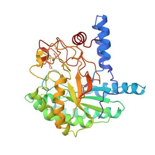
Structural changes of the active site tunnel of Humicola insolens cellobiohydrolase, Cel6A, upon oligosaccharide binding.

Varrot, A.Schulein, M.Davies, G.J.

(1999) Biochemistry 38: 8884-8891

  • DOI: https://doi.org/10.1021/bi9903998
  • Primary Citation of Related Structures:  
    2BVW

  • PubMed Abstract: 

    The mechanisms of crystalline cellulose degradation by cellulases are of paramount importance for the exploitation of these enzymes in applied processes, such as biomass conversion. Cellulases have traditionally been classified into cellobiohydrolases, which are effective in the degradation of crystalline materials, and endoglucanases, which appear to act on "soluble" regions of the substrate. Humicola insolensCel6A (CBH II) is a cellobiohydrolase from glycoside hydrolase family 6 whose native structure has been determined at 1.9 A resolution [Varrot, A., Hastrup, S., Schülein, M., and Davies, G. J. (1999) Biochem. J. 337, 297-304]. Here we present the structure of the catalytic core domain of Humicola insolens cellobiohydrolase II Cel6A in complex with glucose/cellotetraose at 1.7 A resolution. Crystals of Cel6A, grown in the presence of cellobiose, reveal six binding subsites, with a single glucose moiety bound in the -2 subsite and cellotetraose in the +1 to +4 subsites. The complex structure is strongly supportive of the assignment of Asp 226 as the catalytic acid and consistent with proposals that Asp 405 acts as the catalytic base. The structure undergoes several conformational changes upon substrate binding, the most significant of which is a closing of the two active site loops (residues 174-196 and 397-435) with main-chain movements of up to 4.5 A observed. This complex not only defines the polysaccharide-enzyme interactions but also provides the first three-dimensional demonstration of conformational change in this class of enzymes.


  • Organizational Affiliation

    Structural Biology Laboratory, Department of Chemistry, University of York, U.K.


Macromolecules
Find similar proteins by:  (by identity cutoff)  |  3D Structure
Entity ID: 1
MoleculeChains Sequence LengthOrganismDetailsImage
CELLOBIOHYDROLASE II
A, B
362Humicola insolensMutation(s): 0 
EC: 3.2.1.91
UniProt
Find proteins for Q9C1S9 (Humicola insolens)
Explore Q9C1S9 
Go to UniProtKB:  Q9C1S9
Entity Groups  
Sequence Clusters30% Identity50% Identity70% Identity90% Identity95% Identity100% Identity
UniProt GroupQ9C1S9
Sequence Annotations
Expand
  • Reference Sequence
Oligosaccharides

Help

Entity ID: 2
MoleculeChains Length2D Diagram Glycosylation3D Interactions
beta-D-glucopyranose-(1-4)-beta-D-glucopyranose-(1-4)-beta-D-glucopyranose-(1-4)-beta-D-glucopyranose
C
4N/A
Glycosylation Resources
GlyTouCan:  G00025MO
GlyCosmos:  G00025MO
GlyGen:  G00025MO
Entity ID: 3
MoleculeChains Length2D Diagram Glycosylation3D Interactions
beta-D-glucopyranose-(1-4)-beta-D-glucopyranose-(1-4)-alpha-D-glucopyranose
D
3N/A
Glycosylation Resources
GlyTouCan:  G03861VK
GlyCosmos:  G03861VK
GlyGen:  G03861VK
Small Molecules
Ligands 3 Unique
IDChains Name / Formula / InChI Key2D Diagram3D Interactions
NAG
Query on NAG

Download Ideal Coordinates CCD File 
E [auth A],
M [auth B]
2-acetamido-2-deoxy-beta-D-glucopyranose
C8 H15 N O6
OVRNDRQMDRJTHS-FMDGEEDCSA-N
BGC
Query on BGC

Download Ideal Coordinates CCD File 
F [auth A],
N [auth B]
beta-D-glucopyranose
C6 H12 O6
WQZGKKKJIJFFOK-VFUOTHLCSA-N
GOL
Query on GOL

Download Ideal Coordinates CCD File 
G [auth A]
H [auth A]
I [auth A]
J [auth A]
K [auth A]
G [auth A],
H [auth A],
I [auth A],
J [auth A],
K [auth A],
L [auth A],
O [auth B]
GLYCEROL
C3 H8 O3
PEDCQBHIVMGVHV-UHFFFAOYSA-N
Biologically Interesting Molecules (External Reference) 2 Unique
Experimental Data & Validation

Experimental Data

  • Method: X-RAY DIFFRACTION
  • Resolution: 1.70 Å
  • R-Value Free: 0.226 
  • R-Value Work: 0.175 
  • Space Group: P 1 21 1
Unit Cell:
Length ( Å )Angle ( ˚ )
a = 48.56α = 90
b = 154.43β = 119.31
c = 51.04γ = 90
Software Package:
Software NamePurpose
MOSFLMdata reduction
SCALAdata scaling
AMoREphasing
CCP4refinement
CCP4data scaling

Structure Validation

View Full Validation Report


Entry History 

Deposition Data

Revision History  (Full details and data files)

  • Version 1.0: 2000-02-25
    Type: Initial release
  • Version 1.1: 2007-10-16
    Changes: Version format compliance
  • Version 1.2: 2011-07-13
    Changes: Non-polymer description, Version format compliance
  • Version 1.3: 2019-12-25
    Changes: Advisory, Data collection, Derived calculations
  • Version 2.0: 2020-07-29
    Type: Remediation
    Reason: Carbohydrate remediation
    Changes: Advisory, Atomic model, Data collection, Derived calculations, Non-polymer description, Structure summary
  • Version 2.1: 2023-08-23
    Changes: Data collection, Database references, Refinement description, Structure summary