2BIS

Structure of glycogen synthase from Pyrococcus abyssi


Experimental Data Snapshot

  • Method: X-RAY DIFFRACTION
  • Resolution: 2.80 Å
  • R-Value Free: 0.266 
  • R-Value Work: 0.200 
  • R-Value Observed: 0.204 

wwPDB Validation   3D Report Full Report


Ligand Structure Quality Assessment 


This is version 1.3 of the entry. See complete history


Literature

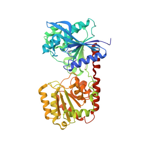
Crystal Structure of an Archaeal Glycogen Synthase: Insights Into Oligomerisation and Substrate Binding of Eukaryotic Glycogen Synthases.

Horcajada, C.Guinovart, J.J.Fita, I.Ferrer, J.C.

(2006) J Biol Chem 281: 2923

  • DOI: https://doi.org/10.1074/jbc.M507394200
  • Primary Citation of Related Structures:  
    2BFW, 2BIS

  • PubMed Abstract: 

    Glycogen and starch synthases are retaining glycosyltransferases that catalyze the transfer of glucosyl residues to the non-reducing end of a growing alpha-1,4-glucan chain, a central process of the carbon/energy metabolism present in almost all living organisms. The crystal structure of the glycogen synthase from Pyrococcus abyssi, the smallest known member of this family of enzymes, revealed that its subunits possess a fold common to other glycosyltransferases, a pair of beta/alpha/beta Rossmann fold-type domains with the catalytic site at their interface. Nevertheless, the archaeal enzyme presents an unprecedented homotrimeric molecular arrangement both in solution, as determined by analytical ultracentrifugation, and in the crystal. The C-domains are not involved in intersubunit interactions of the trimeric molecule, thus allowing for movements, likely required for catalysis, across the narrow hinge that connects the N- and C-domains. The radial disposition of the subunits confers on the molecule a distinct triangular shape, clearly visible with negative staining electron microscopy, in which the upper and lower faces present a sharp asymmetry. Comparison of bacterial and eukaryotic glycogen synthases, which use, respectively, ADP or UDP glucose as donor substrates, with the archaeal enzyme, which can utilize both molecules, allowed us to propose the residues that determine glucosyl donor specificity.


  • Organizational Affiliation

    Departament de Bioquímica i Biologia Molecular, Universitat de Barcelona, Spain.


Macromolecules
Find similar proteins by:  (by identity cutoff)  |  3D Structure
Entity ID: 1
MoleculeChains Sequence LengthOrganismDetailsImage
GLGA GLYCOGEN SYNTHASE
A, B, C
440Pyrococcus abyssiMutation(s): 0 
EC: 2.4.1.21
UniProt
Find proteins for Q9V2J8 (Pyrococcus abyssi (strain GE5 / Orsay))
Explore Q9V2J8 
Go to UniProtKB:  Q9V2J8
Entity Groups  
Sequence Clusters30% Identity50% Identity70% Identity90% Identity95% Identity100% Identity
UniProt GroupQ9V2J8
Sequence Annotations
Expand
  • Reference Sequence
Small Molecules
Ligands 4 Unique
IDChains Name / Formula / InChI Key2D Diagram3D Interactions
UDP
Query on UDP

Download Ideal Coordinates CCD File 
S [auth C]URIDINE-5'-DIPHOSPHATE
C9 H14 N2 O12 P2
XCCTYIAWTASOJW-XVFCMESISA-N
GLC
Query on GLC

Download Ideal Coordinates CCD File 
D [auth A],
R [auth C]
alpha-D-glucopyranose
C6 H12 O6
WQZGKKKJIJFFOK-DVKNGEFBSA-N
GOL
Query on GOL

Download Ideal Coordinates CCD File 
I [auth A]
J [auth A]
K [auth A]
O [auth B]
P [auth B]
I [auth A],
J [auth A],
K [auth A],
O [auth B],
P [auth B],
Q [auth B],
W [auth C]
GLYCEROL
C3 H8 O3
PEDCQBHIVMGVHV-UHFFFAOYSA-N
DIO
Query on DIO

Download Ideal Coordinates CCD File 
E [auth A]
F [auth A]
G [auth A]
H [auth A]
L [auth B]
E [auth A],
F [auth A],
G [auth A],
H [auth A],
L [auth B],
M [auth B],
N [auth B],
T [auth C],
U [auth C],
V [auth C]
1,4-DIETHYLENE DIOXIDE
C4 H8 O2
RYHBNJHYFVUHQT-UHFFFAOYSA-N
Experimental Data & Validation

Experimental Data

  • Method: X-RAY DIFFRACTION
  • Resolution: 2.80 Å
  • R-Value Free: 0.266 
  • R-Value Work: 0.200 
  • R-Value Observed: 0.204 
  • Space Group: C 1 2 1
Unit Cell:
Length ( Å )Angle ( ˚ )
a = 202.985α = 90
b = 73.973β = 131.19
c = 148.221γ = 90
Software Package:
Software NamePurpose
REFMACrefinement
DENZOdata reduction
SCALEPACKdata scaling
MOLREPphasing

Structure Validation

View Full Validation Report


Ligand Structure Quality Assessment 


Entry History 

Deposition Data

Revision History  (Full details and data files)

  • Version 1.0: 2005-11-28
    Type: Initial release
  • Version 1.1: 2011-05-08
    Changes: Version format compliance
  • Version 1.2: 2011-07-13
    Changes: Version format compliance
  • Version 1.3: 2020-07-29
    Type: Remediation
    Reason: Carbohydrate remediation
    Changes: Advisory, Data collection, Derived calculations, Other, Structure summary