The structure of glycogen phosphorylase b with an alkyldihydropyridine-dicarboxylic acid compound, a novel and potent inhibitor.
Zographos, S.E., Oikonomakos, N.G., Tsitsanou, K.E., Leonidas, D.D., Chrysina, E.D., Skamnaki, V.T., Bischoff, H., Goldmann, S., Watson, K.A., Johnson, L.N.(1997) Structure 5: 1413-1425
- PubMed: 9384557
- DOI: https://doi.org/10.1016/s0969-2126(97)00292-x
- Primary Citation of Related Structures:
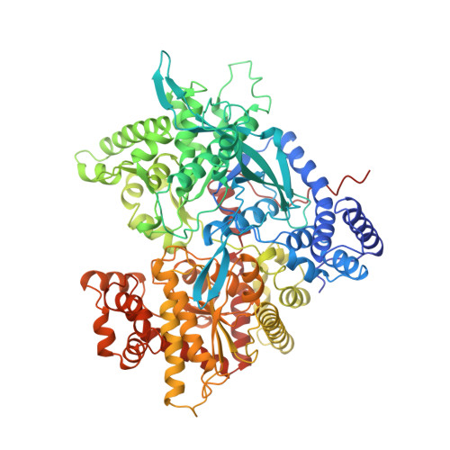
2AMV - PubMed Abstract:
In muscle and liver, glycogen concentrations are regulated by the reciprocal activities of glycogen phosphorylase (GP) and glycogen synthase. An alkyl-dihydropyridine-dicarboxylic acid has been found to be a potent inhibitor of GP, and as such has potential to contribute to the regulation of glycogen metabolism in the non-insulin-dependent diabetes diseased state. The inhibitor has no structural similarity to the natural regulators of GP. We have carried out structural studies in order to elucidate the mechanism of inhibition. Kinetic studies with rabbit muscle glycogen phosphorylase b (GPb) show that the compound (-)(S)-3-isopropyl 4-(2-chlorophenyl)-1,4-dihydro-1-ethyl-2-methyl-pyridine-3,5, 6-tricarboxylate (Bay W1807) has a Ki = 1.6 nM and is a competitive inhibitor with respect to AMP. The structure of the cocrystallised GPb-W1807 complex has been determined at 100K to 2.3 A resolution and refined to an R factor of 0.198 (Rfree = 0.287). W1807 binds at the GPb allosteric effector site, the site which binds AMP, glucose-6-phosphate and a number of other phosphorylated ligands, and induces conformational changes that are characteristic of those observed with the naturally occurring allosteric inhibitor, glucose-6-phosphate. The dihydropyridine-5,6-dicarboxylate groups mimic the phosphate group of ligands that bind to the allosteric site and contact three arginine residues. The high affinity of W1807 for GP appears to arise from the numerous nonpolar interactions made between the ligand and the protein. Its potency as an inhibitor results from the induced conformational changes that lock the enzyme in a conformation known as the T' state. Allosteric enzymes, such as GP, offer a new strategy for structure-based drug design in which the allosteric site can be exploited. The results reported here may have important implications in the design of new therapeutic compounds.
Organizational Affiliation:
Institute of Biological Research and Biotechnology, The National Hellenic Research Foundation 48, vas Constantinou Avenue, Athens, 11635, Greece.