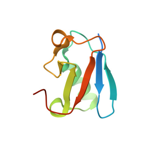
Solution structure and backbone dynamics of an N-terminal ubiquitin-like domain in the GLUT4-regulating protein, TUG.
Tettamanzi, M.C., Yu, C., Bogan, J.S., Hodsdon, M.E.(2006) Protein Sci 15: 498-508
- PubMed: 16501224
- DOI: https://doi.org/10.1110/ps.051901806
- Primary Citation of Related Structures:
2AL3 - PubMed Abstract:
The GLUT4-regulating protein, TUG, functions to retain GLUT4-containing membrane vesicles intracellularly and, in response to insulin stimulation, releases these vesicles to the cellular exocytic machinery for translocation to the plasma membrane. As part of our on going effort to describe the molecular basis for TUG function, we have determined the tertiary structure and characterized the backbone dynamics for an N-terminal ubiquitin-like domain (TUG-UBL1) using NMR spectroscopy. A well-ordered conformation is observed for residues 10-83 of full-length TUG, and confirms a beta-grasp or ubiquitin-like topology. Although not required for in vitro association with GLUT4, the functional role of the TUG-UBL1 domain has not yet been described. We undertook a limited literature review of similar N-terminal UBL domains and noted that a majority participate in protein-protein interactions, generally functioning as adaptor modules to physically associate the over all activity of the protein with a specific cellular process, such as the ubiquitin-proteasome pathway. In consistent fashion, TUG-UBL1 is not expected to participate in a covalent protein modification reaction as it lacks the characteristic C-terminal diglycine ("GG") motif required for conjugation to an acceptor lysine, and also lacks the three most common acceptor lysine residues involved in polyubiquitination. Additionally, analysis of the TUG-UBL1 molecular surface reveals a lack of conservation of the "Ile-44 hydrophobic face" typically involved in ubiquitin recognition. Instead, we speculate on the possible significance of a concentrated area of negative electrostatic potential with increased backbone mobility, both of which are features suggestive of a potential protein-protein interaction site.
- Department of Laboratory Medicine, Yale University, New Haven, CT 06520-8035, USA.
Organizational Affiliation: