Calcium binding in alpha-amylases: an X-ray diffraction study at 2.1-A resolution of two enzymes from Aspergillus.
Boel, E., Brady, L., Brzozowski, A.M., Derewenda, Z., Dodson, G.G., Jensen, V.J., Petersen, S.B., Swift, H., Thim, L., Woldike, H.F.(1990) Biochemistry 29: 6244-6249
- PubMed: 2207069
- DOI: https://doi.org/10.1021/bi00478a019
- Primary Citation of Related Structures:
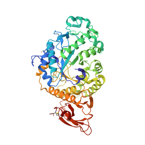
2AAA - PubMed Abstract:
X-ray diffraction analysis (at 2.1-A resolution) of an acid alpha-amylase from Aspergillus niger allowed a detailed description of the stereochemistry of the calcium-binding sites. The primary site (which is essential in maintaining proper folding around the active site) contains a tightly bound Ca2+ with an unusually high number of eight ligands (O delta 1 and O delta 2 of Asp175, O delta of Asn121, main-chain carbonyl oxygens of Glu162 and Glu210, and three water molecules). A secondary binding site was identified at the bottom of the substrate binding cleft; it involves the residues presumed to play a catalytic role (Asp206 and Glu230). This explains the inhibitory effect of calcium observed at higher concentrations. Neutral Aspergillus oryzae (TAKA) alpha-amylase was also refined in a new crystal at 2.1-A resolution. The structure of this homologous (over 80%) enzyme and additional kinetic studies support all the structural conclusions regarding both calcium-binding sites.
- Chemistry Department, University of York, Heslington, U.K.
Organizational Affiliation: