2ZVU

Crystal structure of rat heme oxygenase-1 in complex with ferrous verdoheme


Experimental Data Snapshot

  • Method: X-RAY DIFFRACTION
  • Resolution: 2.20 Å
  • R-Value Free: 0.240 
  • R-Value Work: 0.189 
  • R-Value Observed: 0.192 

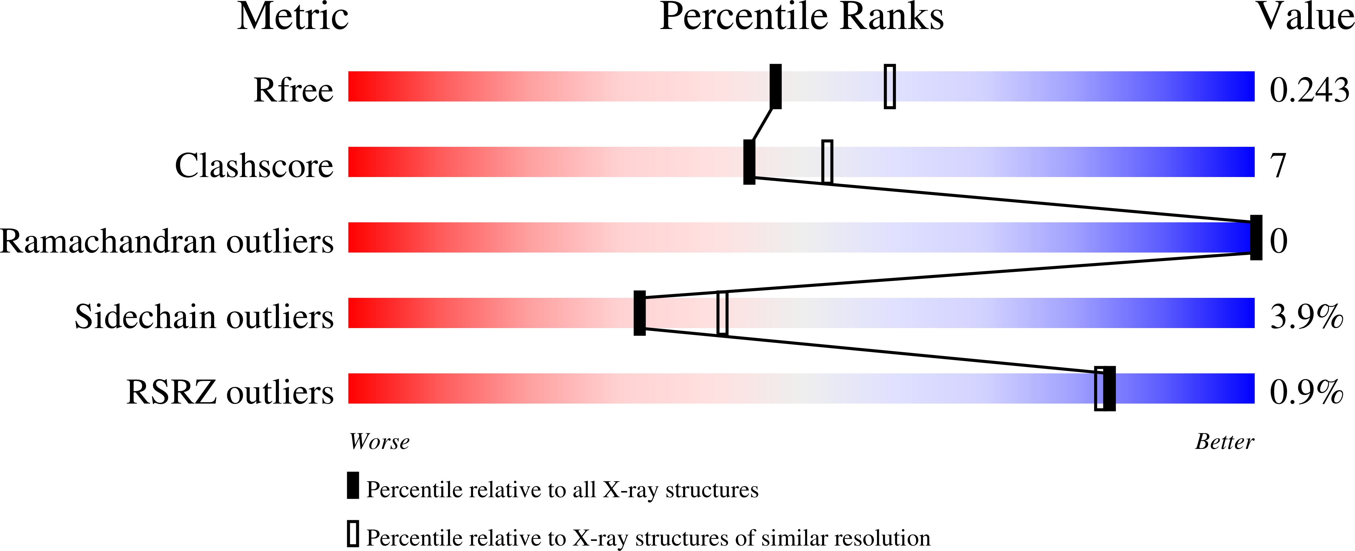
wwPDB Validation   3D Report Full Report


Ligand Structure Quality Assessment 


This is version 1.2 of the entry. See complete history


Literature

Crystal structure of rat haem oxygenase-1 in complex with ferrous verdohaem: presence of a hydrogen-bond network on the distal side

Sato, H.Sugishima, M.Sakamoto, H.Higashimoto, Y.Shimokawa, C.Fukuyama, K.Palmer, G.Noguchi, M.

(2009) Biochem J 419: 339-345

  • DOI: https://doi.org/10.1042/BJ20082279
  • Primary Citation of Related Structures:  
    2ZVU

  • PubMed Abstract: 

    HO (haem oxygenase) catalyses the degradation of haem to biliverdin, CO and ferrous iron via three successive oxygenation reactions, i.e. haem to alpha-hydroxyhaem, alpha-hydroxyhaem to alpha-verdohaem and alpha-verdohaem to ferric biliverdin-iron chelate. In the present study, we determined the crystal structure of ferrous alpha-verdohaem-rat HO-1 complex at 2.2 A (1 A=0.1 nm) resolution. The overall structure of the verdohaem complex was similar to that of the haem complex. Water or OH- was co-ordinated to the verdohaem iron as a distal ligand. A hydrogen-bond network consisting of water molecules and several amino acid residues was observed at the distal side of verdohaem. Such a hydrogen-bond network was conserved in the structures of rat HO-1 complexes with haem and with the ferric biliverdin-iron chelate. This hydrogen-bond network may act as a proton donor to form an activated oxygen intermediate, probably a ferric hydroperoxide species, in the degradation of alpha-verdohaem to ferric biliverdin-iron chelate similar to that seen in the first oxygenation step.


  • Organizational Affiliation

    Department of Medical Biochemistry, Kurume University School of Medicine, 67 Asahi-machi, Kurume 830-0011, Japan.


Macromolecules
Find similar proteins by:  (by identity cutoff)  |  3D Structure
Entity ID: 1
MoleculeChains Sequence LengthOrganismDetailsImage
Heme oxygenase 1267Rattus norvegicusMutation(s): 0 
Gene Names: Hmox1
EC: 1.14.99.3
UniProt
Find proteins for P06762 (Rattus norvegicus)
Explore P06762 
Go to UniProtKB:  P06762
Entity Groups  
Sequence Clusters30% Identity50% Identity70% Identity90% Identity95% Identity100% Identity
UniProt GroupP06762
Sequence Annotations
Expand
  • Reference Sequence
Small Molecules
Ligands 2 Unique
IDChains Name / Formula / InChI Key2D Diagram3D Interactions
VEA
Query on VEA

Download Ideal Coordinates CCD File 
B [auth A]5-OXA-PROTOPORPHYRIN IX CONTAINING FE
C33 H31 Fe N4 O5
OCHHJFVQYXRHAA-HPQJSUICSA-M
FMT
Query on FMT

Download Ideal Coordinates CCD File 
C [auth A]FORMIC ACID
C H2 O2
BDAGIHXWWSANSR-UHFFFAOYSA-N
Experimental Data & Validation

Experimental Data

  • Method: X-RAY DIFFRACTION
  • Resolution: 2.20 Å
  • R-Value Free: 0.240 
  • R-Value Work: 0.189 
  • R-Value Observed: 0.192 
  • Space Group: P 32 2 1
Unit Cell:
Length ( Å )Angle ( ˚ )
a = 65.346α = 90
b = 65.346β = 90
c = 120.24γ = 120
Software Package:
Software NamePurpose
REFMACrefinement
HKL-2000data collection
HKL-2000data reduction
HKL-2000data scaling
MOLREPphasing

Structure Validation

View Full Validation Report


Ligand Structure Quality Assessment 


Entry History 

Deposition Data

Revision History  (Full details and data files)

  • Version 1.0: 2009-02-03
    Type: Initial release
  • Version 1.1: 2011-07-13
    Changes: Version format compliance
  • Version 1.2: 2023-11-01
    Changes: Data collection, Database references, Derived calculations, Refinement description