2Z73

Crystal structure of squid rhodopsin


Experimental Data Snapshot

  • Method: X-RAY DIFFRACTION
  • Resolution: 2.50 Å
  • R-Value Free: 0.206 
  • R-Value Work: 0.188 
  • R-Value Observed: 0.188 

wwPDB Validation   3D Report Full Report


Ligand Structure Quality Assessment 


This is version 1.4 of the entry. See complete history


Literature

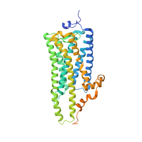
Crystal structure of squid rhodopsin.

Murakami, M.Kouyama, T.

(2008) Nature 453: 363-367

  • DOI: https://doi.org/10.1038/nature06925
  • Primary Citation of Related Structures:  
    2Z73

  • PubMed Abstract: 

    Invertebrate phototransduction uses an inositol-1,4,5-trisphosphate signalling cascade in which photoactivated rhodopsin stimulates a G(q)-type G protein, that is, a class of G protein that stimulates membrane-bound phospholipase Cbeta. The same cascade is used by many G-protein-coupled receptors, indicating that invertebrate rhodopsin is a prototypical member. Here we report the crystal structure of squid (Todarodes pacificus) rhodopsin at 2.5 A resolution. Among seven transmembrane alpha-helices, helices V and VI extend into the cytoplasmic medium and, together with two cytoplasmic helices, they form a rigid protrusion from the membrane surface. This peculiar structure, which is not seen in bovine rhodopsin, seems to be crucial for the recognition of G(q)-type G proteins. The retinal Schiff base forms a hydrogen bond to Asn 87 or Tyr 111; it is far from the putative counterion Glu 180. In the crystal, a tight association is formed between the amino-terminal polypeptides of neighbouring monomers; this intermembrane dimerization may be responsible for the organization of hexagonally packed microvillar membranes in the photoreceptor rhabdom.


  • Organizational Affiliation

    Department of Physics, Graduate School of Science, Nagoya University, Nagoya 464-8602, Japan.


Macromolecules
Find similar proteins by:  (by identity cutoff)  |  3D Structure
Entity ID: 1
MoleculeChains Sequence LengthOrganismDetailsImage
Rhodopsin
A, B
448Todarodes pacificusMutation(s): 0 
Membrane Entity: Yes 
UniProt
Find proteins for P31356 (Todarodes pacificus)
Explore P31356 
Go to UniProtKB:  P31356
Entity Groups  
Sequence Clusters30% Identity50% Identity70% Identity90% Identity95% Identity100% Identity
UniProt GroupP31356
Sequence Annotations
Expand
  • Reference Sequence
Small Molecules
Ligands 6 Unique
IDChains Name / Formula / InChI Key2D Diagram3D Interactions
PC1
Query on PC1

Download Ideal Coordinates CCD File 
N [auth B]1,2-DIACYL-SN-GLYCERO-3-PHOSPHOCHOLINE
C44 H88 N O8 P
NRJAVPSFFCBXDT-HUESYALOSA-N
TWT
Query on TWT

Download Ideal Coordinates CCD File 
G [auth A],
M [auth B]
DOCOSANE
C22 H46
HOWGUJZVBDQJKV-UHFFFAOYSA-N
BOG
Query on BOG

Download Ideal Coordinates CCD File 
C [auth A],
H [auth B]
octyl beta-D-glucopyranoside
C14 H28 O6
HEGSGKPQLMEBJL-RKQHYHRCSA-N
RET
Query on RET

Download Ideal Coordinates CCD File 
D [auth A],
J [auth B]
RETINAL
C20 H28 O
NCYCYZXNIZJOKI-OVSJKPMPSA-N
PLM
Query on PLM

Download Ideal Coordinates CCD File 
E [auth A],
F [auth A],
K [auth B],
L [auth B]
PALMITIC ACID
C16 H32 O2
IPCSVZSSVZVIGE-UHFFFAOYSA-N
SO4
Query on SO4

Download Ideal Coordinates CCD File 
I [auth B]SULFATE ION
O4 S
QAOWNCQODCNURD-UHFFFAOYSA-L
Experimental Data & Validation

Experimental Data

  • Method: X-RAY DIFFRACTION
  • Resolution: 2.50 Å
  • R-Value Free: 0.206 
  • R-Value Work: 0.188 
  • R-Value Observed: 0.188 
  • Space Group: P 62
Unit Cell:
Length ( Å )Angle ( ˚ )
a = 122.55α = 90
b = 122.55β = 90
c = 158.74γ = 120
Software Package:
Software NamePurpose
ADSCdata collection
MOLREPphasing
CNSrefinement
MOSFLMdata reduction
SCALAdata scaling

Structure Validation

View Full Validation Report


Ligand Structure Quality Assessment 


Entry History 

Deposition Data

Revision History  (Full details and data files)

  • Version 1.0: 2008-05-13
    Type: Initial release
  • Version 1.1: 2011-07-13
    Changes: Version format compliance
  • Version 1.2: 2017-10-11
    Changes: Data collection
  • Version 1.3: 2020-07-29
    Type: Remediation
    Reason: Carbohydrate remediation
    Changes: Data collection, Derived calculations, Structure summary
  • Version 1.4: 2023-11-01
    Changes: Data collection, Database references, Refinement description, Structure summary