2YZ3

Crystallographic Investigation of Inhibition Mode of the VIM-2 Metallo-beta-lactamase from Pseudomonas aeruginosa with Mercaptocarboxylate Inhibitor


Experimental Data Snapshot

  • Method: X-RAY DIFFRACTION
  • Resolution: 2.30 Å
  • R-Value Free: 0.247 
  • R-Value Work: 0.212 
  • R-Value Observed: 0.212 

wwPDB Validation   3D Report Full Report


This is version 1.2 of the entry. See complete history


Literature

Crystallographic investigation of the inhibition mode of a VIM-2 metallo-beta-lactamase from Pseudomonas aeruginosa by a mercaptocarboxylate inhibitor.

Yamaguchi, Y.Jin, W.Matsunaga, K.Ikemizu, S.Yamagata, Y.Wachino, J.Shibata, N.Arakawa, Y.Kurosaki, H.

(2007) J Med Chem 50: 6647-6653

  • DOI: https://doi.org/10.1021/jm701031n
  • Primary Citation of Related Structures:  
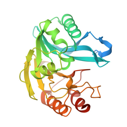
    2YZ3

  • PubMed Abstract: 

    The VIM-2 metallo-beta-lactamase enzyme from Pseudomonas aeruginosa catalyzes the hydrolysis of most beta-lactam antibiotics including carbapenems, and there are currently no potent inhibitors of such enzymes. We found rac-2-omega-phenylpropyl-3-mercaptopropionic acid, phenylC3SH, to be a potent inhibitor of VIM-2. The structure of the VIM-2-phenylC3SH complex was determined by X-ray crystallography to 2.3 A. The structure revealed that the thiol group of phenylC3SH bridged to the two zinc(II) ions and the phenyl group interacted with Tyr67(47) on loop1 near the active site, by pi-pi stacking interactions. The methylene group interacted with Phe61(42) located at the bottom of loop1 through CH-pi interactions. Dynamic movements were observed in Arg228(185) and Asn233(190) on loop2, compared with the native structure (PDB code: 1KO3 ). These results suggest that the above-mentioned four residues play important roles in the binding and recognition of inhibitors or substrates and in stabilizing a loop in the VIM-2 enzyme.


  • Organizational Affiliation

    Environmental Safety Center, Kumamoto University, Department of Structure-Function Physical Chemistry, Graduate School of Pharmaceutical Sciences, Japan. yyamagu@gpo.kumamoto-u.ac.jp


Macromolecules
Find similar proteins by:  (by identity cutoff)  |  3D Structure
Entity ID: 1
MoleculeChains Sequence LengthOrganismDetailsImage
Metallo-beta-lactamase
A, B
266Pseudomonas putidaMutation(s): 0 
Gene Names: VIM-2bla VIM-2blaVIM-2
EC: 3.5.2.6
UniProt
Find proteins for Q7BJM5 (Pseudomonas putida)
Explore Q7BJM5 
Go to UniProtKB:  Q7BJM5
Entity Groups  
Sequence Clusters30% Identity50% Identity70% Identity90% Identity95% Identity100% Identity
UniProt GroupQ7BJM5
Sequence Annotations
Expand
  • Reference Sequence
Small Molecules
Binding Affinity Annotations 
IDSourceBinding Affinity
M5P PDBBind:  2YZ3 Ki: 220 (nM) from 1 assay(s)
Binding MOAD:  2YZ3 Ki: 220 (nM) from 1 assay(s)
Experimental Data & Validation

Experimental Data

  • Method: X-RAY DIFFRACTION
  • Resolution: 2.30 Å
  • R-Value Free: 0.247 
  • R-Value Work: 0.212 
  • R-Value Observed: 0.212 
  • Space Group: P 21 21 21
Unit Cell:
Length ( Å )Angle ( ˚ )
a = 45.184α = 90
b = 90.749β = 90
c = 128.977γ = 90
Software Package:
Software NamePurpose
CNSrefinement
HKL-2000data collection
HKL-2000data reduction
HKL-2000data scaling
SOLVEphasing

Structure Validation

View Full Validation Report


Entry History 

Deposition Data

Revision History  (Full details and data files)

  • Version 1.0: 2008-03-11
    Type: Initial release
  • Version 1.1: 2011-07-13
    Changes: Version format compliance
  • Version 1.2: 2024-03-13
    Changes: Data collection, Database references, Derived calculations