2X06

SULFOLACTATE DEHYDROGENASE FROM METHANOCALDOCOCCUS JANNASCHII


Experimental Data Snapshot

  • Method: X-RAY DIFFRACTION
  • Resolution: 2.50 Å
  • R-Value Free: 0.224 
  • R-Value Work: 0.153 
  • R-Value Observed: 0.157 

wwPDB Validation   3D Report Full Report


Ligand Structure Quality Assessment 


This is version 1.5 of the entry. See complete history


Literature

Methanoarchaeal Sulfolactate Dehydrogenase: Prototype of a New Family of Nadh-Dependent Enzymes.

Irimia, A.Madern, D.Vellieux, F.M.D.

(2004) EMBO J 23: 1234

  • DOI: https://doi.org/10.1038/sj.emboj.7600147
  • Primary Citation of Related Structures:  
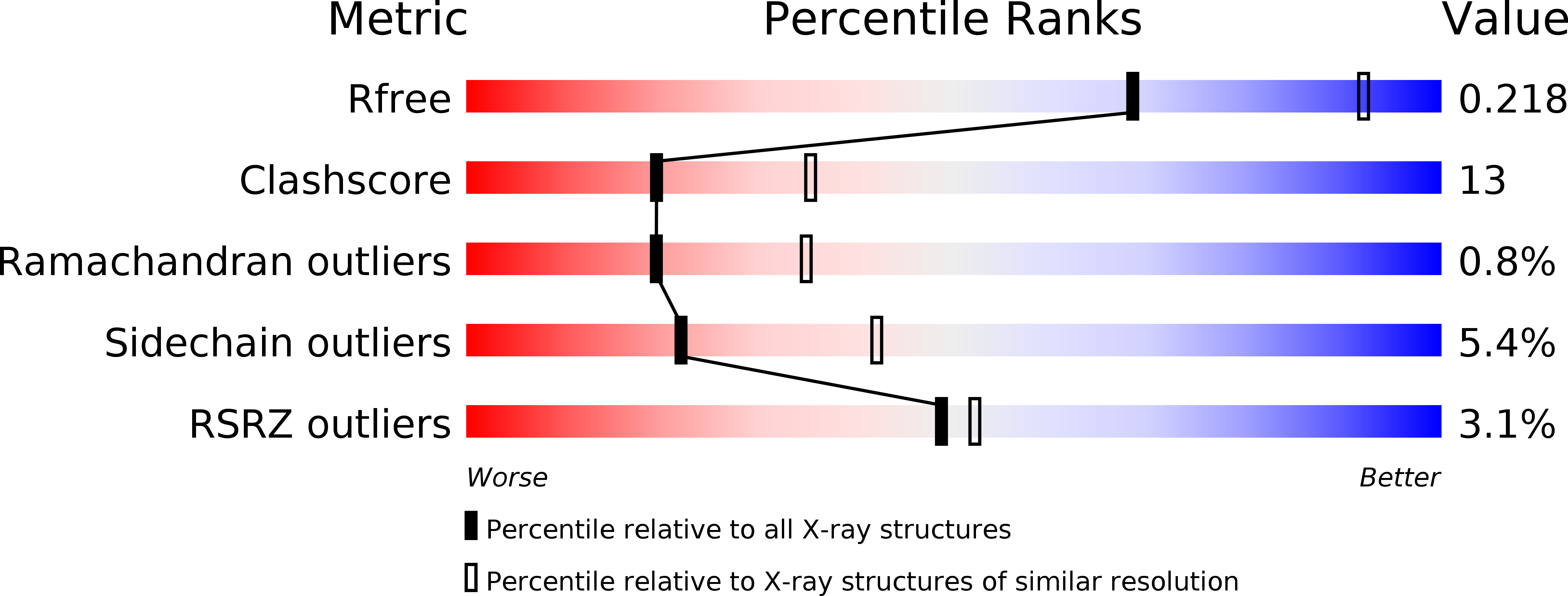
    2X06

  • PubMed Abstract: 

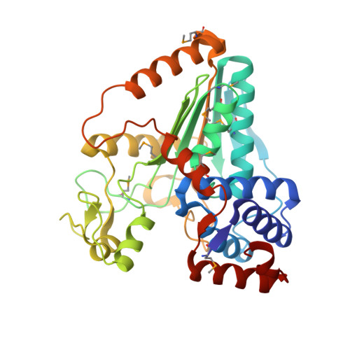
    The crystal structure of the sulfolactate dehydrogenase from the hyperthermophilic and methanogenic archaeon Methanocaldococcus jannaschii was solved at 2.5 A resolution (PDB id. 1RFM). The asymmetric unit contains a tetramer of tight dimers. This structure, complexed with NADH, does not contain a cofactor-binding domain with 'Rossmann-fold' topology. Instead, the tertiary and quaternary structures indicate a novel fold. The NADH is bound in an extended conformation in each active site, in a manner that explains the pro-S specificity. Cofactor binding involves residues belonging to both subunits within the tight dimers, which are therefore the smallest enzymatically active units. The protein was found to be a homodimer in solution by size-exclusion chromatography, analytical ultracentrifugation and small-angle neutron scattering. Various compounds were tested as putative substrates. The results indicate the existence of a substrate discrimination mechanism, which involves electrostatic interactions. Based on sequence homology and phylogenetic analyses, several other enzymes were classified as belonging to this novel family of homologous (S)-2-hydroxyacid dehydrogenases.


  • Organizational Affiliation

    Laboratoire de Biophysique Moléculaire, Institut de Biologie Structurale J-P Ebel CEA CNRS UJF, Grenoble, France.


Macromolecules
Find similar proteins by:  (by identity cutoff)  |  3D Structure
Entity ID: 1
MoleculeChains Sequence LengthOrganismDetailsImage
L-SULFOLACTATE DEHYDROGENASE
A, B, C, D, E
A, B, C, D, E, F, G, H
344Methanocaldococcus jannaschiiMutation(s): 0 
EC: 1.1.1.272
UniProt
Find proteins for Q58820 (Methanocaldococcus jannaschii (strain ATCC 43067 / DSM 2661 / JAL-1 / JCM 10045 / NBRC 100440))
Explore Q58820 
Go to UniProtKB:  Q58820
Entity Groups  
Sequence Clusters30% Identity50% Identity70% Identity90% Identity95% Identity100% Identity
UniProt GroupQ58820
Sequence Annotations
Expand
  • Reference Sequence
Experimental Data & Validation

Experimental Data

  • Method: X-RAY DIFFRACTION
  • Resolution: 2.50 Å
  • R-Value Free: 0.224 
  • R-Value Work: 0.153 
  • R-Value Observed: 0.157 
  • Space Group: P 1 21 1
Unit Cell:
Length ( Å )Angle ( ˚ )
a = 80.93α = 90
b = 203.71β = 112.05
c = 100.05γ = 90
Software Package:
Software NamePurpose
XDSdata reduction
XSCALEdata scaling
TRUNCATEdata scaling
SnBphasing
MLPHAREphasing
PHENIXrefinement

Structure Validation

View Full Validation Report


Ligand Structure Quality Assessment 


Entry History 

Deposition Data

Revision History  (Full details and data files)

  • Version 1.0: 2009-12-15
    Type: Initial release
  • Version 1.1: 2011-05-08
    Changes: Version format compliance
  • Version 1.2: 2011-07-13
    Changes: Version format compliance
  • Version 1.3: 2017-07-05
    Changes: Refinement description
  • Version 1.4: 2019-01-30
    Changes: Data collection, Experimental preparation
  • Version 1.5: 2019-02-06
    Changes: Data collection, Experimental preparation