2V8M

Carbohydrate-binding of the starch binding domain of Rhizopus oryzae glucoamylase in complex with beta-cyclodextrin and maltoheptaose


Experimental Data Snapshot

  • Method: X-RAY DIFFRACTION
  • Resolution: 2.30 Å
  • R-Value Free: 0.280 
  • R-Value Work: 0.238 
  • R-Value Observed: 0.238 

wwPDB Validation   3D Report Full Report


This is version 2.0 of the entry. See complete history


Literature

Crystal Structures of the Starch-Binding Domain from Rhizopus Oryzae Glucoamylase Reveal a Polysaccharide-Binding Path.

Tung, J.-Y.Chang, M.D.-T.Chou, W.-I.Liu, Y.-Y.Yeh, Y.Chang, F.Lin, S.Qiu, Z.Sun, Y.-J.

(2008) Biochem J 416: 27

  • DOI: https://doi.org/10.1042/BJ20080580
  • Primary Citation of Related Structures:  
    2V8L, 2V8M, 2VQ4

  • PubMed Abstract: 

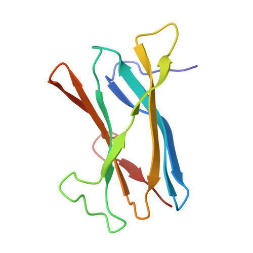
    GA (glucoamylase) hydrolyses starch and polysaccharides to beta-D-glucose. RoGA (Rhizopus oryzae GA) consists of two functional domains, an N-terminal SBD (starch-binding domain) and a C-terminal catalytic domain, which are connected by an O-glycosylated linker. In the present study, the crystal structures of the SBD from RoGA (RoGACBM21) and the complexes with beta-cyclodextrin (SBD-betaCD) and maltoheptaose (SBD-G7) were determined. Two carbohydrate binding sites, I (Trp(47)) and II (Tyr(32)), were resolved and their binding was co-operative. Besides the hydrophobic interaction, two unique polyN loops comprising consecutive asparagine residues also participate in the sugar binding. A conformational change in Tyr(32) was observed between unliganded and liganded SBDs. To elucidate the mechanism of polysaccharide binding, a number of mutants were constructed and characterized by a quantitative binding isotherm and Scatchard analysis. A possible binding path for long-chain polysaccharides in RoGACBM21 was proposed.


  • Organizational Affiliation

    Institute of Bioinformatics and Structural Biology, National Tsing Hua University, Hsinchu 30013, Taiwan.


Macromolecules
Find similar proteins by:  (by identity cutoff)  |  3D Structure
Entity ID: 1
MoleculeChains Sequence LengthOrganismDetailsImage
GLUCOAMYLASE A
A, B, C, D
106Rhizopus arrhizusMutation(s): 0 
EC: 3.2.1.3
UniProt
Find proteins for B7XC04 (Rhizopus oryzae)
Explore B7XC04 
Go to UniProtKB:  B7XC04
Entity Groups  
Sequence Clusters30% Identity50% Identity70% Identity90% Identity95% Identity100% Identity
UniProt GroupB7XC04
Sequence Annotations
Expand
  • Reference Sequence
Oligosaccharides

Help

Entity ID: 2
MoleculeChains Length2D Diagram Glycosylation3D Interactions
alpha-D-glucopyranose-(1-4)-alpha-D-glucopyranose-(1-4)-alpha-D-glucopyranose-(1-4)-alpha-D-glucopyranose-(1-4)-alpha-D-glucopyranose-(1-4)-alpha-D-glucopyranose-(1-4)-alpha-D-glucopyranose
E, F, G, H
7N/A
Glycosylation Resources
GlyTouCan:  G41288IQ
GlyCosmos:  G41288IQ
GlyGen:  G41288IQ
Experimental Data & Validation

Experimental Data

  • Method: X-RAY DIFFRACTION
  • Resolution: 2.30 Å
  • R-Value Free: 0.280 
  • R-Value Work: 0.238 
  • R-Value Observed: 0.238 
  • Space Group: P 1 21 1
Unit Cell:
Length ( Å )Angle ( ˚ )
a = 37.686α = 90
b = 110.908β = 90.72
c = 61.161γ = 90
Software Package:
Software NamePurpose
CNSrefinement
HKL-2000data reduction
HKL-2000data scaling
MOLREPphasing

Structure Validation

View Full Validation Report


Entry History 

Deposition Data

Revision History  (Full details and data files)

  • Version 1.0: 2008-08-19
    Type: Initial release
  • Version 1.1: 2012-02-22
    Changes: Database references, Other, Version format compliance
  • Version 2.0: 2020-07-29
    Type: Remediation
    Reason: Carbohydrate remediation
    Changes: Advisory, Atomic model, Data collection, Derived calculations, Other, Structure summary